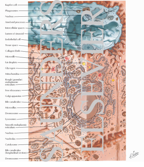
Ultramicroscopic Structure of Normal Liver Cells
Image ID: 12857
Pricing
Good For All Uses:
Print, Electronic and Digital
Please Note: Specialized programs, such as Adobe Illustrator, are needed to use EPS files.
Print, Electronic and Digital
Please Note: Specialized programs, such as Adobe Illustrator, are needed to use EPS files.
The preview images do not contain enough pixels to make small type legible on screen, however,
the actual delivered images will contain labels that are clearly legible. (view PDF preview)
Other Versions of This Illustration

This Illustration was Published In
Netter's Essential Histology
Author: William K. Ovalle, Patrick C. Nahirney
Chapter: Liver
Page: 319
Author: William K. Ovalle, Patrick C. Nahirney
Chapter: Liver
Page: 319
The Netter Collection of Medical Illustrations - Digestive System, Part III - Liver, Biliary Tract and Pancreas
Author: Frank H. Netter, MD
Chapter: Structure, Metabolism, Diagnostic, Surgical Procedures of Liver Diseases
Page: 170
Author: Frank H. Netter, MD
Chapter: Structure, Metabolism, Diagnostic, Surgical Procedures of Liver Diseases
Page: 170
Netter's Histology Flash Cards
Author: William K. Ovalle and Patrick C. Nahirney
Chapter: Liver, Gallbladder, and Exocrine Pancreas
Author: William K. Ovalle and Patrick C. Nahirney
Chapter: Liver, Gallbladder, and Exocrine Pancreas
If you can see this message, you need to update your Flash Player.

