
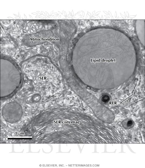
Electron Micrograph of Part of a Hepatocyte Showing Sagittal and Cross-Sectional Smooth Endoplasmic Reticulum
ID: 12996
Title:
Electron Micrograph of Part of a Hepatocyte Showing Sagittal and Cross-Sectional Smooth Endoplasmic Reticulum
Category:
Labeled - Ovalle Histology 1E
Please describe! how you will use this image and then you will be able to add this image to your shopping basket.


 Please Wait...