
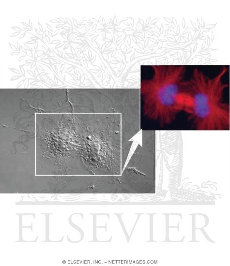
Light Micrographs of Cultured Cells Showing Events of Mitosis: Anaphase
ID: 13092
Title:
Light Micrographs of Cultured Cells Showing Events of Mitosis: Anaphase
Category:
Labeled - Ovalle Histology 1E
Please describe! how you will use this image and then you will be able to add this image to your shopping basket.


 Please Wait...