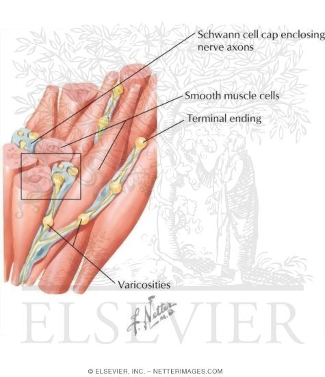
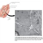
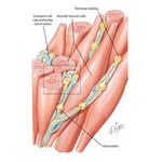
Electron Micrograph of Smooth Muscle Cells Close to Nerve Axons In Transverse Section
Image ID: 13390
Pricing
EM of smooth muscle cells close to nerve axons in transverse section. Several smooth muscle cells (Sm) are in the field of view; one is sectioned at the level of its nucleus (N). A group of unmyelinated axons (Ax) supported by the process of a Schwann cell (SC) is seen in surrounding interstitial connective tissue. An axonal varicosity contains a cluster of synaptic vesicles (SV). 37,000×.
The preview images do not contain enough pixels to make small type legible on screen, however,
the actual delivered images will contain labels that are clearly legible. (view PDF preview)
Other Versions of This Illustration

This Illustration was Published In
Netter's Essential Histology
Author: William K. Ovalle, Patrick C. Nahirney
Chapter: Muscle Tissue
Page: 99
Author: William K. Ovalle, Patrick C. Nahirney
Chapter: Muscle Tissue
Page: 99
If you can see this message, you need to update your Flash Player.
©2005–2012 Elsevier. All Rights Reserved.


