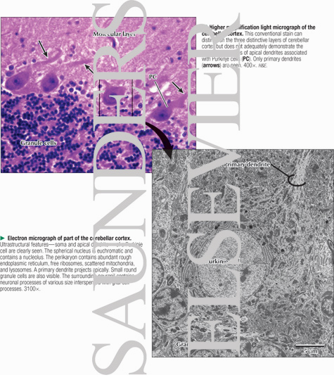
Higher Magnification Light Micrograph of the Cerebellar Cortex With Electron Micrograph of Part of the Cerebellar Cortex
Image ID: 13445
Pricing
Higher magnification LM of the cerebellar cortex. This conventional stain can distinguish the three distinctive layers of cerebellar cortex but does not adequately demonstrate the extensive branches of apical dendrites associated with Purkinje cells (PC). Only primary dendrites (arrows) are seen. 400×. H&E.
EM of part of the cerebellar cortex. Ultrastructural features—soma and apical dendrite—of a Purkinje cell are clearly seen. The spherical nucleus is euchromatic and contains a nucleolus. The perikaryon contains abundant rough endoplasmic reticulum, free ribosomes, scattered mitochondria, and lysosomes. A primary dendrite projects apically. Small round granule cells are also visible. The surrounding neuropil contains neuronal processes of various size interspersed with glial cell processes. 3100×.
The preview images do not contain enough pixels to make small type legible on screen, however,
the actual delivered images will contain labels that are clearly legible. (view PDF preview)
This Illustration was Published In
Netter's Essential Histology
Author: William K. Ovalle, Patrick C. Nahirney
Chapter: Nervous Tissue
Page: 122
Author: William K. Ovalle, Patrick C. Nahirney
Chapter: Nervous Tissue
Page: 122
If you can see this message, you need to update your Flash Player.
©2005–2012 Elsevier. All Rights Reserved.


