
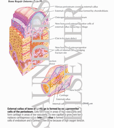
Bone Repair (Intermediate Phase)
ID: 13554
Title:
Bone Repair (Intermediate Phase)
Category:
Labeled - Ovalle Histology 1E
Please describe! how you will use this image and then you will be able to add this image to your shopping basket.


 Please Wait...