
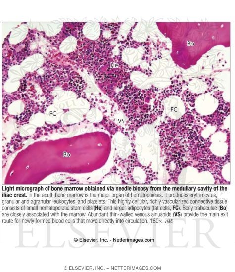
Light Micrograph of Bone Marrow Obtained Via Needle Biopsy From the Medullary Cavity of the Iliac Crest
ID: 13567
Title:
Light Micrograph of Bone Marrow Obtained Via Needle Biopsy From the Medullary Cavity of the Iliac Crest
Category:
Labeled - Ovalle Histology 1E
Please describe! how you will use this image and then you will be able to add this image to your shopping basket.


 Please Wait...