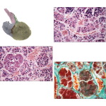
Light Micrograph of the Anterior Lobe Showing Chromophils and Chromophobes and Tinctorial Differences Between Chromophobes and the Two Kinds of Chromophils
Image ID: 13999
Pricing
LM of chromophils and chromophobes in the anterior lobe. Cytoplasm of chromophils (arrows) is stained; that of smaller chromophobes is not, but their nuclei (arrowheads) are. Cells are interspersed with sinusoidal capillaries (Cap) and delicate connective tissue stroma. Parenchymal cells are round to polygonal. Chromophobes usually have small, heterochromatic nuclei; chromophils have larger, euchromatic nuclei with prominent nucleoli. 420×. H&E.
LM of the anterior lobe. The different staining pattern of the small acidophils and larger basophils reflects their granule content. A large network of sinusoidal capillaries (Cap) is between the clumps of parenchymal cells. The vessels receive hormones released by these cells and deliver releasing or inhibiting factors from the hypothalamohypophyseal portal system to affect cells of the anterior lobe. 420×. H&E.
The preview images do not contain enough pixels to make small type legible on screen, however,
the actual delivered images will contain labels that are clearly legible. (view PDF preview)
If you can see this message, you need to update your Flash Player.
©2005–2012 Elsevier. All Rights Reserved.


