
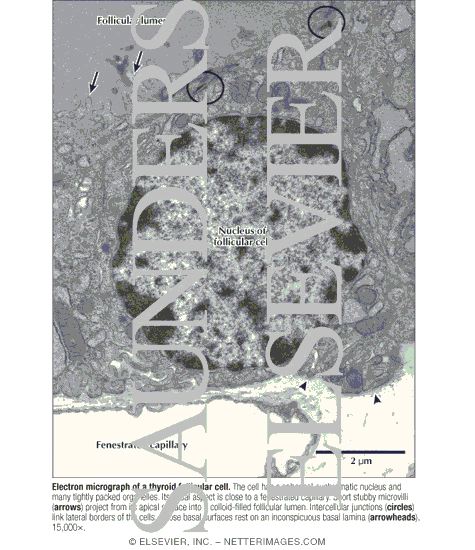
Electron Micrograph of a Thyroid Follicular Cell
ID: 14022
Category: Labeled
Please describe! how you will use this image and then you will be able to add this image to your shopping basket.
ID: 14022
Category: Labeled


 Please Wait...