
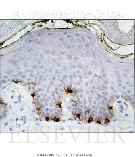
Immunostained Light Micrograph of Thick Skin Showing Melanocytes In the Epidermis
ID: 14229
Category: Unlabeled - Ovalle Histology 1E
Please describe! how you will use this image and then you will be able to add this image to your shopping basket.


 Please Wait...