
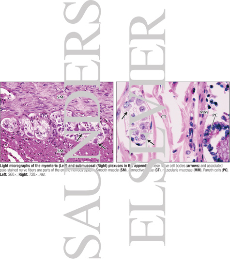
Light Micrographs of the Myenteric (Left) and Submucosal (Right) Plexuses In the Appendix
ID: 14424
Category: Labeled
Please describe! how you will use this image and then you will be able to add this image to your shopping basket.
ID: 14424
Category: Labeled


 Please Wait...