
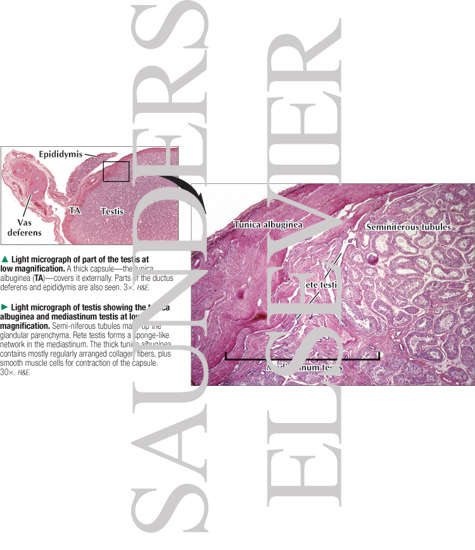
Light Micrograph of Part of the Testis at Low Magnification With Light Micrograph of Testis Showing the Tunica Albuginea and Mediastinum Testis at Low Magnification
ID: 14644
Category: Labeled
Please describe! how you will use this image and then you will be able to add this image to your shopping basket.


 Please Wait...