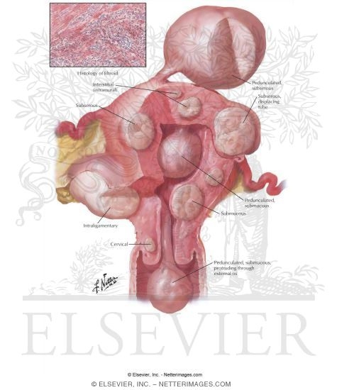
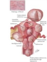
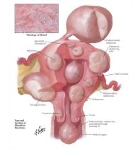
Myoma (Fibroid) I - Locations
Image ID: 5170
Pricing
Good For All Uses:
Print, Electronic and Digital
Please Note: Specialized programs, such as Adobe Illustrator, are needed to use EPS files.
Print, Electronic and Digital
Please Note: Specialized programs, such as Adobe Illustrator, are needed to use EPS files.
The preview images do not contain enough pixels to make small type legible on screen, however,
the actual delivered images will contain labels that are clearly legible. (view PDF preview)
This Illustration was Published In
Netter's Clinical Anatomy
Author: John T. Hansen, PhD and David R. Lambert, MD
Chapter: Pelvis and Perineum
Page: 482
Author: John T. Hansen, PhD and David R. Lambert, MD
Chapter: Pelvis and Perineum
Page: 482
Netter's Illustrated Human Pathology
Author: Maximilian L. Buja, MD and Gerhard R. F. Krueger
Chapter: Diseases of the Female Reproductive System
Page: 281
Author: Maximilian L. Buja, MD and Gerhard R. F. Krueger
Chapter: Diseases of the Female Reproductive System
Page: 281
Netter's Obstetrics, Gynecology and Women's Health
Author: Roger P. Smith, MD
Chapter: Uterine Pathology
Page: 140
Author: Roger P. Smith, MD
Chapter: Uterine Pathology
Page: 140
+ view/hide full list of publications
The Netter Collection of Medical Illustrations - Reproductive System
Author: Frank H. Netter, MD
Chapter: Diseases of the Uterus
Page: 166
Author: Frank H. Netter, MD
Chapter: Diseases of the Uterus
Page: 166
If you can see this message, you need to update your Flash Player.


