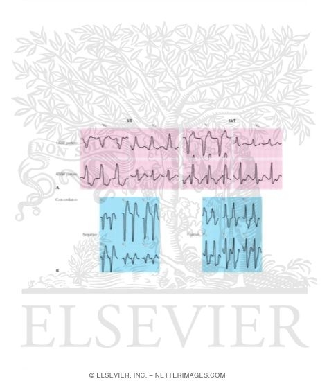
Figure 29-2 Changes in QRS morphology in ventricular tachycardia (VT) and in supraventricular tachycardia (SVT). (A) Typical patterns seen with left bundle branch block (LBBB) and in right bundle branch block (RBBB). (B) Typical patterns of positive and negative concordance in QRS complexes. See text for details.
The preview images do not contain enough pixels to make small type legible on screen, however,
the actual delivered images will contain labels that are clearly legible.
This Illustration was Published In
Netter's Cardiology
Author: Marschall S. Runge, George A. Stouffer, Cam Patterson
Chapter: Ventricular Tachycardia
Page: 243
Author: Marschall S. Runge, George A. Stouffer, Cam Patterson
Chapter: Ventricular Tachycardia
Page: 243
If you can see this message, you need to update your Flash Player.
©2005–2012 Elsevier. All Rights Reserved.


