
Stable Fracture Vertebral Dislocation and Fractures
ID: 8072
Title:
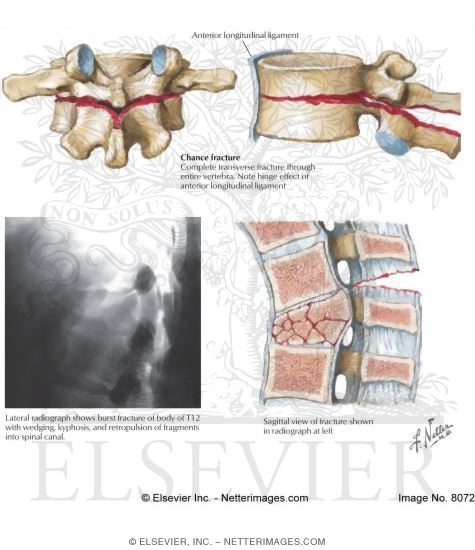
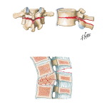
Vertebral Dislocation and Fractures
Category:
Labeled - Hansen Clinical Anatomy 1E
Please describe! how you will use this image and then you will be able to add this image to your shopping basket.


 Please Wait...