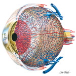
Vascular Supply of Eye
Image ID: 8815
Pricing
Good For All Uses:
Print, Electronic and Digital
Please Note: Specialized programs, such as Adobe Illustrator, are needed to use EPS files.
Print, Electronic and Digital
Please Note: Specialized programs, such as Adobe Illustrator, are needed to use EPS files.
Labels in This Image
- Vascular arrangements within the choroid (vascular tunic) of the eyeball
- Anterior tributaries of a vorticose vein
- Anterior ciliary arteries
- Episcleral vein*
- Tendon of superior rectus muscle
- Junctions of suprachoroidal
- tributaries forming vorticose vein
- Sclera
- Superior ophthalmic vein
- Posterior conjunctival
- artery and vein
- Bulb of vorticose (choroid) vein
- Vorticose (choroid) vein
- Anterior ciliary veins
- Long posterior ciliary artery
- Major arterial circle of iris
- Short posterior
- ciliary arteries
- Minor arterial
- circle of iris
- External sheath of
- optic nerve (dura mater)
- Conjunctiva
- and cornea
- Internal sheath
- of optic nerve
- (pia-arachnoid mater)
- Short posterior
- ciliary arteries
- Central retinal
- Arteries and
- artery and vein
- veins of iris
- Scleral venous sinus
- Vessels of internal
- (canal of Schlemm)
- sheath of optic nerve
- Veins draining scleral venous
- sinus (into anterior ciliary veins)
- Short posterior ciliary arteries
- Anterior ciliary veins
- Segment of episcleral vein*
- Nonpigmented and pigmented
- regions of the retina
- Muscular artery
- Inferior ophthalmic vein
- Recurrent branch of anterior ciliary artery
- Posterior tributaries of a vorticose vein
- *The episcleral veins are shown here anastomosing with the vorticose veins,
- which they do; however they also drain into the anterior ciliary veins.
The preview images do not contain enough pixels to make small type legible on screen, however,
the actual delivered images will contain labels that are clearly legible. (view labels, view PDF preview)
This Illustration was Published In
If you can see this message, you need to update your Flash Player.


