
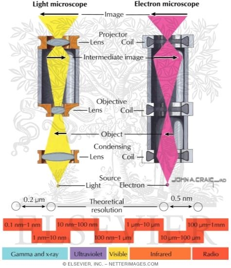
Optical Parts of a Conventional Light Microscope Or Transmission Electron Microscope (TEM)
ID: 12921
Title:
Optical Parts of a Conventional Light Microscope Or Transmission Electron Microscope (TEM)
Category:
Labeled - Ovalle Histology 1E
Please describe! how you will use this image and then you will be able to add this image to your shopping basket.


 Please Wait...