
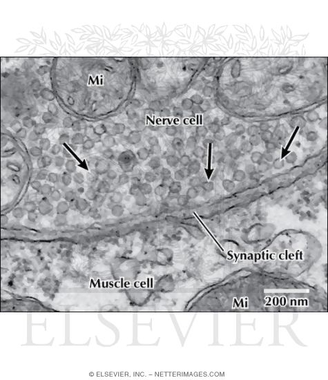
Electron Micrograph of Synaptic Vesicles at a Neuromuscular Junction
ID: 13070
Title:
Electron Micrograph of Synaptic Vesicles at a Neuromuscular Junction
Category:
Labeled - Ovalle Histology 1E
Please describe! how you will use this image and then you will be able to add this image to your shopping basket.


 Please Wait...