
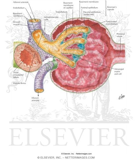
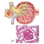
Histology of Renal Corpuscle
ID: 8970
Title:
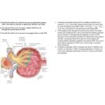
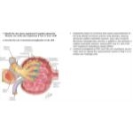
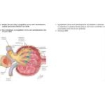
Anatomy of the Glomerulus
Category:
Labeled - Hansen Atlas Physiology 1E
Please describe! how you will use this image and then you will be able to add this image to your shopping basket.


 Please Wait...