
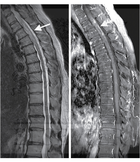
(A) Sagittal T2 MRI of the thoracic spinal cord showing extensive abnormal T2 hyperintense signal in the lower cervical and upper thoracic cord with more than three segments involved, characteristic of NMO. (B) Sagittal T1 Fat Sat postcontrast image demonstrating peripheral intramedullary enhancement from T2 through T4
ID: 77719
Category: Unlabeled
Please describe! how you will use this image and then you will be able to add this image to your shopping basket.


 Please Wait...