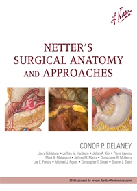
Surgical Anatomy - Delaney
Author: Conor P. Delaney
ISBN: 9781437708332
The Neck
- Page 5: Patient Positioning and Anatomy in Neck Dissection
- Page 7: Flap Elevation in Neck Dissection
- Page 9: Dissection of Level I (Submental and Submandibular Regions)
- Page 11: Dissection of Levels II (Upper Jugular Modal Chain) and III (Midjugular Nodal Chain)
- Page 12: Lymphadenectomy (Levels I-III)
Endocrine
- Page 23: Thyroid Gland, Anterior View
- Page 25: Anatomic Landmarks for Thyroidectomy or Parathyroidectomy Incision
- Page 27: Surgical Anatomy for Thyroidectomy and Parathyroidectomy
- Page 29: Anatomy of Superior and Recurrent Laryngeal Nerves
- Page 31: Preoperative Imaging of Neck: Scintigram and Sonogram
- Page 33: Anatomy and Embryology of Parathyroid Glands
- Page 35: Anatomic Sites for Ectopic Parathyroid Adenoma, With Images of Abnormal Focus and A Corresponding Specimen
- Page 38: Adrenal Glands in Situ, Anterior Views
- Page 39: Histology of Adrenal Glands
- Page 41: Port Site Placement for Transabdominal Laparoscopic Adrenalectomy
- Page 43: Peritoneum of Posterior Abdominal Wall
- Page 44: Right Laparoscopic Adrenalectomy Technique
- Page 45: Arteries and Veins of Suprarenal Glands in Situ.
- Page 47: Left Laparoscopic Adrenalectomy Technique
Upper Gastrointestinal
- Page 52: Esophagus in Situ
- Page 53: Indications for Esophagectomy
- Page 55: Anatomy of the Lesser Curve, Second Portion of the Duodenum, and Gastrohepatic Ligament (Stomach is Rotated Cephalad)
- Page 57: Transhiatal Esophagectomy
- Page 59: Ivor Lewis Esophagectomy: Five-Port Laparascopic Placement
- Page 61: Modified McKeown Esophagectomy: Azygos System of Veins and Esophageal Lymph Nodes/Vessels
- Page 63: Subdivisions of Mediastinum and Cardiac Nerves
- Page 67: Arteries of Esophagus
- Page 68: Veins of Esophagus
- Page 69: Innervation of Esophagus
- Page 71: Sliding Hiatal Hernia
- Page 73: Anatomy for Esophageal Mobilization
- Page 75: Anatomy for Esophageal Mobilization, Continued
- Page 77: Anatomy for Gastric Mobilization
- Page 79: Laparoscopic Nissen (360-degree) Fundoplication and Endoscopy
- Page 80: Crural Closure, Nissen (360-degree) Fundoplication, and Gastropexy
- Page 83: Complications of Gastric and Duodenal Ulcers
- Page 84: Arterial Supply of Stomach
- Page 85: Endoscopic and Upper Gastrointestinal Evaluation
- Page 87: Innervation and Arterial Supply of Stomach
- Page 88: Innervation of Stomach and Duodenum
- Page 89: Vagal Control of Gastric Secretion
- Page 91: Truncal and Highly Selective Vagotomies
- Page 95: Stomach in Situ
- Page 96: Arterial Supply of Stomach
- Page 97: Venous and Lymphatic Drainage of Stomach
- Page 99: Distal and Proximal Gastric Cancer and Reconstruction
- Page 103: Duodenal Bulb, Blood Supply, and Endoscopic View
- Page 105: Duodenal Ulcers
- Page 106: Anatomy of Structures Adjacent to Duodenum
- Page 107: Pyloroplasty Constructions
Hepatobiliary
- Page 129: Gallbladder and Extrahepatic Bile Ducts and Arterial Supply
- Page 130: Variations in Cystic and Hepatic Ducts
- Page 131: Hepatic Artery Variations
- Page 133: Cholelithiasis (Gallstones)
- Page 135: Cholecystectomy and the Critical View
- Page 137: Intraoperative Imaging in Cholecystectomy
- Page 139: Postcholecystectomy Syndromes
- Page 143: Cystic Duct Anatomy and Variants
- Page 145: Choledocholithiasis: Pathologic Features
- Page 147: Transystic and Transductal/Choledochotomy Approaches
- Page 149: Open Common Bile Duct Exploration
- Page 151: Choledochoduodenostomy
- Page 155: Liver Segments and Lobes: Vessel and Duct Distribution
- Page 156: Variations in Origin and Course of Hepatic Artery and Branches
- Page 157: Hepatic Artery: Magnetic Resonance Image and Surgical View
- Page 159: Variations in Cystic and Hepatic Ducts
- Page 160: Variations in Form of Liver
- Page 161: Cirrhosis I: Pathways of Formation
- Page 163: Right Hepatic Lobectomy
- Page 165: Right Hepatic Lobectomy, Continued
- Page 167: Left Hepatic Lobectomy
- Page 169: Left Hepatic Lobectomy, Continued
- Page 173: Anatomy for Preoperative Evaluation of Pancreas
- Page 175: Open Retrograde Distal Pancreatectomy With Splenectomy
- Page 177: Division of Splenic Artery and Vein and Pancreas
- Page 179: Lymphatic Drainage of Pancreas
- Page 181: Mobilization of Pancreas During Spleen-Preserving Distal Pancreatectomy
- Page 183: Port Site Placement for Laparoscopic Distal Pancreatectomy
- Page 187: Pancreatic Cancer: Clinical Features
- Page 189: Arterial Supply of Stomach and Duodenum
- Page 190: Variations in Origin and Course of Hepatic Artery and Branches
- Page 191: Hepatic Portal Vein Tributaries: Portocaval Anastomoses
- Page 193: Liver and Arterial Anatomy of Hepatoduodenal Ligament for Suprapancreatic Dissection
- Page 195: Venous Anatomy and the Resection Bed
- Page 199: Spleen and Surrounding Structures
- Page 201: Venous Anatomy and the Resection Bed in Splenectomy
- Page 205: Abdominal Anatomy and Organ Procurement Exposures
- Page 207: Suprahepatic Aortic Exposure During Abdominal Organ Procurement
- Page 209: Pancreas and Kidney Procurement
- Page 211: Anatomic Variations of Kidney Allograft
- Page 213: Vascular Anastomoses of Kidney Allograft
- Page 215: Ureteral Anastomosis of Kidney Allograft
- Page 217: Arterial Reconstruction for Pancreas Allograft
- Page 219: Superior Mesenteric Vein Anatomy for Portal Drainage of Pancreas Allograft
- Page 221: End-Stage Liver Disease
- Page 223: Liver Anatomy
- Page 225: Liver Surgical Approaches and Completed Transplant
- Page 227: Anastomoses in Liver Transplant
Lower Gastrointestinal
- Page 233: Autonomic Innervation of the Intestine
- Page 235: Cross-Sectional Periappendicular Anatomy
- Page 237: Ileocecal Region
- Page 239: Vermiform Appendix
- Page 241: Variations in Cecal and Appendicular Arteries
- Page 242: Ileocecal Region and Iliac Vessels
- Page 243: Right Lower Quadrant Anatomy: Laparoscopic View
- Page 247: Abdominal Wall Anatomy and Rectus Sheath
- Page 249: Ileostomy: Anatomic Landmarks and Surgical Technique
- Page 251: End Colostomy
- Page 253: Brooke Ileostomy Technique
- Page 255: Loop-End Iliostomy Technique
- Page 259: Right Colectomy and Arteries of the Colon
- Page 261: Retroperitoneal Structures
- Page 263: Variations in Vascular Anatomy of Right Colon
- Page 267: Left Colectomy: Authors' Preference for Laparoscopic Port Placement
- Page 269: Vascular Supply to Colon
- Page 270: Left Resection Pattern; Autonomic Nerves and Lymphatic Drainage to Colon
- Page 271: Vascular Variations in Conon; Duodenal Anatomy
- Page 273: Splenic Flexure
- Page 275: Left Hemicolectomy: Skeletonization
- Page 279: Hepatic and Splenic Flexures
- Page 281: Greater Omental and Stomach in Transverse Colectomy
- Page 283: Arteries of Large Intestines
- Page 285: Middle Colic Artery in Transverse Colectomy
- Page 289: Pelvic Viscera and Perineum: Female
- Page 290: Endopelvic Fascia and Potential Spaces: Female
- Page 291: Arteries and Veins of Colon and Rectum
- Page 292: Autonomic Nerves and Ganglia of Abdomen
- Page 293: Anatomic Relations of Ureters: Male and Female
- Page 295: Inferior Mesenteric Artery
- Page 297: Inferior Mesenteric Vein and Splenic Flexure
- Page 299: Upper Posterior Mesorectal Dissection
- Page 301: Lower Anterior Mesorectal Dissection
- Page 303: Stapled Rectal Transection
- Page 305: Intersphincteric Transanal Transection of Rectum
- Page 309: Proctoscopy and Endorectal Ultrasonography
- Page 311: Arteries and Nerves of Pelvis and Rectum
- Page 313: Approach for Rectal Dissection
- Page 315: Prostate and Seminal Vesicles
- Page 317: Perineal Dissection
- Page 321: Anatomy and Types of Hemorrhoids
- Page 323: Office Procedures for Hemorrhoids
- Page 325: Hemorrhoidectomy and Incarcerated (Strangulated) Hemorrhoids
- Page 329: Sites of Periectal Abscess
- Page 331: Perianal Innervation and Patient Positioning
- Page 333: Deep Postanal Space Abscess
- Page 335: Types of Anorectal Fistula and Goodsail's Rule
- Page 337: Perirectal Fistula Examination and Management
Hernia
- Page 343: Inguinal Region: Dissections
- Page 344: Patent Processus Vaginalis and Indirect Inguinal Hernia
- Page 345: Types of Hernias: Incarcerated, Strangulated, and Amyand's (Appendix in Sac)
- Page 347: Abdominal Wall and Anatomic Landmarks in Hernia Repair
- Page 349: Exposed Anatomy for Hernia Repair
- Page 351: Tension-Free Hernia Repair
- Page 353: Bassini and McVay Hernia Repairs
- Page 357: Anterior and Posterior Views of Myopectineal Orifice
- Page 359: Posterior and Anterior Views of Inguinal Region
- Page 361: Inguinal Landmarks in Hernia Repair: Warning Triangles and Corona Mortis
- Page 363: Laparoscopic Dissection: Transabdominal Preperitoneal (TAPP) Approach to Inguinal Hernia
- Page 365: Laparoscopic Balloon Dissection: Totally Extraperitoneal (TEP) Approach to Inguinal Hernia
- Page 369: Anatomy of Femoral Hernia
- Page 371: Open Surgical Repair of Femoral Hernia
- Page 373: Laparoscopic Surgical Repair of Femoral Hernia
- Page 377: Innervation of Abdomen and Perineum
- Page 378: Course and Relations of Intercostal Nerves and Arteries
- Page 379: Arteries of Anterior Abdominal Wall
- Page 381: Cross Section of Anterior Abdominal Wall and Creation of Retrorectus Space
- Page 383: Posterior View of Pelvis and Lower Abdominal Wall
- Page 385: Open Ventral (Abdominal) Hernia Repair
Vascular
- Page 401: Abdominal Incision Lines and Exposure of Midline Retroperitoneum
- Page 403: Aortic and Lilac Arterial Relationships With Retroperitoneal Structures
- Page 405: Aortic Relationships: Colon, Duodenum, and Left Renal Vein
- Page 407: Aortic Relationships With Retroperitoneum and Left Renal Vein
- Page 409: Aortic Relationships With Lumbar Spine and Visceral Vessels
- Page 411: Abdominal Aortic Aneurysm and Anatomy
- Page 451: Arteries of Knee and Thigh Incisions
- Page 452: Muscles of Thigh: Anterior View
- Page 453: Muscles of Knee and Thigh (Deep Dissection): Medial and Anterior Views
- Page 455: Muscles of Thigh (Superficial/Deeper Dissections) and Leg (Superficial Dissection)
- Page 456: Popiliteal Artery Aneurysm Before (left) and After (right) PTFE Bypass Graft
- Page 459: Fascial Compartment of Leg
- Page 460: Cross-Sectional Anatomy of Thigh
- Page 461: Arteries and Veins of Leg
- Page 463: Below-Knee Amputation and Closure
- Page 465: Above-Knee Amputation and Closure
Vascular Access and Emergency Procedures
- Page 479: Arrangement of Tendons, Vessels, and Nerves at the Wrist
- Page 481: Axilla (Dissection: Anterior View) and Brachial Artery in Situ
- Page 483: Arteries and Nerves of Thigh: Deep Dissection (Anterior View)
- Page 484: Muscles, Arteries, and Nerves of Front of Ankle and Dorsum of Foot: Deeper Dissection
- Page 487: Cross-Sectional Anatomy of Leg and Fascial Compartments
- Page 488: Cross-Sectional Leg Anatomy, Below Knee
- Page 489: Cross-Sectional Leg Anatomy, Middle and Lower Tibia
- Page 491: Etiology of Compartment Syndrome
- Page 493: Clinical Diagnosis of Compartment Syndrome
- Page 494: Common Fibular (Peroneal) Nerve: Mixed Motor/Sensory Function
- Page 495: Measurement of Intracompartmental Pressure
- Page 497: Incisions for Compartment Syndrome of Leg
- Page 499: Muscles of Leg With Superficial Peroneal Nerve: Lateral View
- Page 501: Individual Muscles of Forearm: Flexors and Extensors
- Page 503: Nerves of Upper Limb
- Page 505: Cross-Sectional Anatomy and Incisions for Compartment Syndrome of Forearm and Hand
- Page 506: Median and Ulnar Nerve in Forearm
- Page 509: Anatomy of Thorax
- Page 511: Lung Topography, Lungs in Situ, and Chest Wall Cross Section
- Page 513: Landmarks for Chest Tube Placement
- Page 527: Saggital Section of Pharynx
- Page 529: Views of Larynx
- Page 531: Nose and Lower Airway/Trachea
- Page 533: Indications and Examination for Intubation
- Page 535: Laryngoscopy
- Page 537: Sinus Endoscopy
Breast and Oncology
- Page 543: Partial Mastectomy
- Page 545: Total Mastectomy With Modified Radical Technique
- Page 549: Management Algorithm for Nipple Discharge
- Page 551: Duct Excision and Intraductal Papilloma
- Page 555: Sentinel Lymph Node Biopsy
- Page 557: Axillary, Cervical, Popliteal, and Inguinal Lymph Nodes
- Page 559: Identification for ""Hot"" Lymph Node
- Page 563: Axilla (Dissection) and Lymph Vessels/Nodes of Mammary Gland
- Page 565: Axillary Lymph Node Dissection
- Page 567: Inguinal Lymph Node Dissection
Urology and Gynecology
- Page 579: Uterus, Ovaries, and Uterine Tubes
- Page 580: Ligamentous and Fascial Support of Pelvic Viscera
- Page 581: Arteries and Veins of Pelvic Organs and Ureteral Injury
- Page 583: Abdominal Hysterectomy: Isolation of Round and Infundibulopelvic Ligaments
- Page 585: Pelvic Cross Section With Peritoneum Removed
- Page 609: Robot-assisted Prostatectomy and Surgical Approaches to Prostate
- Page 611: Posterior Prostatic Dissection
- Page 613: Development of Space Retzius
- Page 615: Bladder Neck Dissection and Neurovascular Bundle
- Page 617: Prostatic Pedicle Ligation and Division of Deep Dorsal Venous Plexus
- Page 619: Division of Urethra and Vesicourethral Anastomosis
- Page 623: Male: Fascial Planes and Pelvic Contents
- Page 625: Peritoneum of Posterior Abdominal Wall
- Page 627: Lymph Vessels and Nodes of Kidneys and Bladder
- Page 629: Male: Vascular Supply of Pelvic Organs and Loss of Erection
- Page 631: Pelvic Viscera and Perineum: Male
- Page 633: Female: Vascular Supply of Pelvic Organs and Anterior Exenteration Resections
- Page 635: Urethra Incision and Resection

