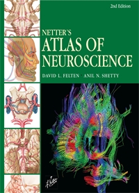
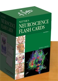
Flash Cards - Neuroscience, Felten 2E
2nd Edition
Author: David L. Felten
ISBN: 9781437709407
- Page 1.01: Neuronal Structure
- Page 1.02: Neuronal Cell Types
- Page 1.03: Glial Cell Types
- Page 1.04: The Blood-Brain Barrier
- Page 1.05: Myelination of CNS and PNS Axons
- Page 1.06: Synaptic Morphology
- Page 1.07: Conduction Velocity
- Page 1.08: Foramina in the Base of the Adult Skull
- Page 1.09: Bony Framework of the Head and Neck
- Page 1.1: Schematic of the Meninges and Their Relationships to the Brain and Skull
- Page 1.11: Hematomas
- Page 1.12: Surface Anatomy of the Forebrain: Lateral View
- Page 1.13: Lateral View of the Forebrain: Functional Regions
- Page 1.14: Anatomy of the Medial (Midsagittal) Surface of the Brain In Situ
- Page 1.15: Anatomy of the Basal Surface of the Brain, with the Brain Stem and Cerebellum Removed
- Page 1.16: Brain Imaging: Computed Tomography Scans, Coronal and Sagittal
- Page 1.17: Brain Imaging: Magnetic Resonance Imaging, Axial and Sagittal T1-Weighted Images
- Page 1.18: Brain Imaging: Magnetic Resonance Imaging, Axial and Sagittal T1-Weighted Images
- Page 1.19: Horizontal Brain Sections Showing the Basal Ganglia
- Page 1.2: Major Limbic Forebrain Structures
- Page 1.21: Color Imaging of the Corpus Callosum by Diffusion Tensor Imaging
- Page 1.22: Hippocampal Formation and Fornix
- Page 1.23: Thalamic Anatomy
- Page 1.24: Thalamic Nuclei
- Page 1.25: Brain Stem Surface Anatomy: Posterolateral View
- Page 1.26: Brain Stem Surface Anatomy: Anterior View
- Page 1.27: Cerebellar Anatomy: Internal Features
- Page 1.28: Spinal Column: Bony Anatomy
- Page 1.29: Spinal Cord: Gross Anatomy In Situ
- Page 1.3: The Spinal Cord: Its Meninges and Spinal Roots
- Page 1.31: Spinal Cord: Cross Sectional Anatomy In Situ
- Page 1.32: Spinal Cord White and Gray Matter
- Page 1.33: Ventricular Anatomy
- Page 1.34: Ventricular Anatomy in Coronal Forebrain Section
- Page 1.35: Anatomy of the Fourth Ventricle: Lateral View
- Page 1.36: Magnetic Resonance Imaging of the Ventricles: Axial and Coronal Views
- Page 1.37: Circulation of the Cerebrospinal Fluid
- Page 1.38: Arterial Supply to the Brain and Meninges
- Page 1.39: Arterial Distribution to the Brain: Basal View
- Page 1.4: Arterial Distribution to the Brain: Cutaway Basal View Showing the Circle of Willis
- Page 1.41: Arterial Distribution to the Brain: Coronal Forebrain Section
- Page 1.42: Circle of Willis: Schematic Illustration and Vessels In Situ
- Page 1.43: Arterial Distribution to the Brain: Laterial and Medial Views
- Page 1.44: Magnetic Resonance Angiography: Coronal Full Vessel View
- Page 1.45: Vertebrobasilar Arterial System
- Page 1.46: Arterial Blood Supply to the Spinal Cord: Longitudinal View
- Page 1.47: Arterial Supply to the Spinal Cord: Cross Sectional View
- Page 1.48: Meninges and Superficial Cerebral Veins
- Page 1.49: Venous Sinuses
- Page 1.5: Deep Venous Drainage of the Brain: Relationship to the Ventricles
- Page 1.51: Magnetic Resonance Venography
- Page 1.52: Neurulation
- Page 1.53: Neural Tube Development and Neural Crest Formation
- Page 1.54: Development of Peripheral Axons
- Page 1.55: Early Brain Development: The 36-Day-Old Embryo
- Page 1.56: Early Brain Development: The 49-Day-Old Embryo and the 3-Month-Old Embryo
- Page 1.57: Development of the Eye and Orbit
- Page 1.58: Development of the Ear
- Page 1.59: Development of the Pituitary Gland
- Page 1.6: Development of the Ventricles
- Page 2.001: Spinal Cord and PNS Schematic
- Page 2.002: Anatomy of a Peripheral Nerve
- Page 2.003: Cutaneous Receptors
- Page 2.004: Neuromuscular Junction
- Page 2.005: Dermatomal Distribution
- Page 2.006: Cutaneous Nerves of the Head and Neck
- Page 2.007: Cervical Plexus
- Page 2.008: Phrenic Nerve
- Page 2.009: Thoracic Nerves
- Page 2.01: Brachial Plexus
- Page 2.011: Dermatomes of the Upper Limb
- Page 2.012: Cutaneous Innervation of the Upper Limb from Peripheral Nerves
- Page 2.013: The Scapular, Axillary, and Radial Nerves Above the Elbow
- Page 2.014: Radial Nerve in the Forearm
- Page 2.015: Musculocutaneous Nerve
- Page 2.016: Median Nerve
- Page 2.017: Ulnar Nerve
- Page 2.018: Lumbar Plexus
- Page 2.019: Sacral and Coccygeal Plexuses
- Page 2.02: Femoral and Lateral Femoral Cutaneous Nerves
- Page 2.021: Obturator Nerve
- Page 2.022: Sciatic and Posterior Femoral Cutaneous Nerves
- Page 2.023: Tibial Nerve
- Page 2.024: Common Peroneal Nerve
- Page 2.025: Schematic of the Autonomic Nervous System
- Page 2.026: Autonomic Distribution to the Head and Neck: Medial View
- Page 2.027: Autonomic Distribution to the Head and Neck: Lateral View
- Page 2.028: Autonomic Distribution to the Eye
- Page 2.029: Schematic of the Pterygopalatine and Submandibular Ganglia
- Page 2.03: Thoracic Sympathetic Chain and Splanchnic Nerves
- Page 2.031: Innervation of the Tracheobronchial Tree
- Page 2.032: Innervation of the Heart
- Page 2.033: Abdominal Nerves and Ganglia
- Page 2.034: Nerves of the Esophagus
- Page 2.035: Nerves of the Stomach and Duodenum
- Page 2.036: Nerves of the Small Intestine
- Page 2.037: Nerves of the Large Intestine
- Page 2.038: Enteric Nervous System: Cross Sectional View
- Page 2.039: Autonomic Innervation of the Liver and Biliary Tract
- Page 2.04: Autonomic Innervatin of the Pancreas
- Page 2.041: Schematic of Innervation of the Adrenal Gland
- Page 2.042: Autonomic Pelvic Nerves and Ganglia
- Page 2.043: Neres of the Kidneys, Ureters, and Urinary Bladder
- Page 2.044: Innervation of the Male Reproductive Organs
- Page 2.045: Innervation of the Female Reproductive Organs
- Page 2.046: Cytoarchitecture of the Spinal Cord Gray Matter
- Page 2.047: C7 Spinal Cord Cross Section
- Page 2.048: T8 Spinal Cord Cross Section
- Page 2.049: L3 Spinal Cord Cross Section
- Page 2.05: S3 Spinal Cord Cross Section
- Page 2.051: Spinal Cord Imaging
- Page 2.052: Spinal Somatic Reflex Pathways
- Page 2.053: Muscle and Joint Receptors and Muscle Spindles
- Page 2.054: Brain Stem Cross Section: Medulla-Spinal Cord Transition
- Page 2.055: Brain Stem Cross Section: Medulla at the Level of the Obex
- Page 2.056: Brain Stem Cross Section: Medulla at the Level of the Inferior Olive
- Page 2.057: Brain Stem Cross Section: Medulla at the Level of CN X and the Vestibular Nuclei
- Page 2.058: Brain Stem Cross Section: Medullo-Pontine Junction
- Page 2.059: Brain Stem Cross Section: Pons at the Level of the Facial Nucleus
- Page 2.06: Brain Stem Cross Section: Pons at the Level of the Genu of the Facial Nerve
- Page 2.061: Brain Stem Cross Sections: Pons at the Level of the Trigeminal Motor and Main Sensory Nuclei
- Page 2.062: Brain Stem Cross Section: Pons-Midbrain Junction
- Page 2.063: Brain Stem Cross Section: Midbrain at the Level of the Inferior Colliculus
- Page 2.064: Brain Stem Cross Section: Midbrain at the Level of the Superior Colliculus and Geniculate Nuclei
- Page 2.065: Brain Stem Cross Section: Midbrain-Diencephalic Junction
- Page 2.066: Cranial Nerves: Basal View of the Brain
- Page 2.067: Cranial Nerves and Their Nuclei: Schematic View From Above
- Page 2.068: Nerves of the Orbit
- Page 2.069: Extraocular Cranial Nerves
- Page 2.07: Trigeminal Nerve (CN V)
- Page 2.071: Facial Nerve (CN VII)
- Page 2.072: Vestibulocochlear Nerve (CN VIII)
- Page 2.073: Glossopharyngeal Nerve (CN IX)
- Page 2.074: Vagus Nerve (CN X)
- Page 2.075: Accessory Nerve (CN XI)
- Page 2.076: Hypoglossal Nerve (CN XII)
- Page 2.077: Reticular Formation and Nuclei
- Page 2.078: Sleep-Wakefulness Control
- Page 2.079: Cerebellar Organization: Lobes and Regions
- Page 2.08: Cerebellar Anatomy: Deep Nuclei and Cerebellar Peduncles
- Page 2.081: Thalamic Anatomy and Interconnections with the Cerebral Cortex
- Page 2.082: Hypothalamus and Pituitary Gland
- Page 2.083: Hypothalamic Nuclei
- Page 2.084: Axial Sections Through the Forebrain: Midpons
- Page 2.085: Axial Sections Through the Forebrain: Midbrain
- Page 2.086: Axial Sections Through the Forebrain: Rostral Midbrain and Hypothalamus
- Page 2.087: Axial Sections Through the Forebrain: Anterior Commissure and Caudal Thalamus
- Page 2.088: Axial Sections Through the Forebrain: Head of the Caudate Nucleus and Midthalamus
- Page 2.089: Axial Sections through the Forebrain: Basal Ganglia and Internal Capsule
- Page 2.09: Axial Section through the Forebrain: Dorsal Caudate Nucleus, Splenium and Genu of the Internal Capsule
- Page 2.091: Coronal Sections Through the Forebrain: Genu of Corpus Callosum
- Page 2.092: Coronal Sections Through the Forebrain: Head of the Caudate Nucleus and Nucleus Accumbens
- Page 2.093: Coronal Sections Through the Forebrain: Anterior Commissure and Columns of Fornix
- Page 2.094: Coronal Sections Through the Forebrain: Amygdala, Anterior Limb of Internal Capsule
- Page 2.095: Coronal Sections Through the Forebrain: Mammillary Body
- Page 2.096: Coronal Sections Through the Forebrain: Midthalamus
- Page 2.097: Coronal Sections Through the Forebrain: Geniculate Nuclei
- Page 2.098: Coronal Sections Through the Forebrain: Caudal Pulvinar and Superior Colliculus
- Page 2.099: Cortical Association Pathways
- Page 2.1: Major Cortical Association Bundles
- Page 2.101: Color Imaging of Projection Pathways from the Cerebral Cortex
- Page 2.102: Noradrenergic Pathways
- Page 2.103: Serotonergic Pathways
- Page 2.104: Dopaminergic Pathways
- Page 2.105: Central Cholinergic Pathways
- Page 2.106: Olfactory Nerves
- Page 3.01: Somatosensory Afferents to the Spinal Cord
- Page 3.02: Somatosensory System: Spinocerebellar Pathways
- Page 3.03: Somatosensory System: The Dorsal Column System and Epicritic Modalities
- Page 3.04: Somatosensory System: The Spinothalamic and Spinoreticular Systems and Protopathic Modalities
- Page 3.05: Mechanisms of Neuropathic Pain and Sympathetically Maintained Pain
- Page 3.06: Descending Control of Ascending Somatosensory Systems
- Page 3.07: Trigeminal Sensory and Associated Sensory Systems
- Page 3.08: Taste Pathways
- Page 3.09: Peripheral Pathways for Sound Reception
- Page 3.1: Bony and Membranous Labyrinths
- Page 3.11: VIII Nerve Innervation of Hair Cells of the Organ of Corti
- Page 3.12: Afferent Auditory Pathways
- Page 3.13: Centrifugal (Efferent) Auditory Pathways
- Page 3.14: Vestibular Receptors
- Page 3.15: Vestibular Pathways
- Page 3.16: Nystagmus
- Page 3.17: Anatomy of the Eye
- Page 3.18: Anterior and Posterior Chambers of the Eye
- Page 3.19: The Retina: Retinal Layers
- Page 3.2: Arteries and Veins of the Eye
- Page 3.21: Anatomy and Relations of Optic Chiasm
- Page 3.22: Visual Pathways: Retinal Projections to the Thalamus, Hypothalamus, and Brain Stem
- Page 3.23: Visual Pathway: The Retino-Geniculo-Calcarine Pathway
- Page 3.24: Visual Pathways in the Parietal and Temporal Lobes
- Page 3.25: Distribution of Lower Motor Neurons in the Brain Stem
- Page 3.26: Cortical Efferent Pathways
- Page 3.27: Color Imaging of Cortical Efferent Pathways
- Page 3.28: Corticobulbar Tract
- Page 3.29: Corticospinal Tract
- Page 3.3: Rubrospinal Tract
- Page 3.31: Vestibulospinal Tracts
- Page 3.32: Reticulospinal and Corticoreticular Pathways
- Page 3.33: Central Control of Eye Movements
- Page 3.34: Central Control of Respiration
- Page 3.35: Cerebellar Neuronal Circuitry
- Page 3.36: Afferent Pathways to the Cerebellum
- Page 3.37: Cerebellar Efferent Pathways
- Page 3.38: Connections of the Basal Ganglia
- Page 3.39: General Organization of the Autonomic Nervous System
- Page 3.4: Sections Through the Hypothalamus: Preoptic and Supraoptic Zones
- Page 3.41: Sections Through the Hypothalamus: Tuberal Zone
- Page 3.42: Sections Through the Hypothlamus: Mammillary Zone
- Page 3.43: Schematic Reconstruction of the Hypothalamus
- Page 3.44: Afferent and Efferent Pathways Associated with the Hypothalamus
- Page 3.45: Paraventricular Nucleus of the Hypothalamus: Regulation of Pituitary Neurohormonal Outflow, Autonomic Preganglionic Outflow, and Limbic Activity
- Page 3.46: Mechanisms of Cytokine Influences on the Hypothalamus and Other Brain Regions and on Behavior
- Page 3.47: Circumventricular Organs
- Page 3.48: The Hypophyseal Portal Vasculature
- Page 3.49: Regulation of Anterior Pituitary Hormone Secretion
- Page 3.5: Posterior Pituitary (Neurohypophyseal) Hormones: Oxytocin and Vasopressin
- Page 3.51: The Hypothalamus and Thermoregulation
- Page 3.52: Neuroimmunomodulation
- Page 3.53: Anatomy of the Limbic Forebrain
- Page 3.54: Hippocampal Formation: General Anatomy
- Page 3.55: Neuronal Connections in the Hippocampal Formation
- Page 3.56: Major Afferent and Efferent Connections of the Hippocampal Formation
- Page 3.57: Major Afferent Connections of the Amygdala
- Page 3.58: Major Connections of the Cingulate Cortex
- Page 3.59: Olfactory Pathways

