Anatomy Atlas - 4E
Author: Frank H. Netter
ISBN: 9781416033851
- Page 1: Head and Neck
- Page 2: Skull: Anterior View
- Page 3: Skull: Anteroposterior Radiograph
- Page 4: Skull: Lateral View
- Page 5: Skull: Lateral Radiograph
- Page 6: Skull: Midsagittal Section
- Page 7: Calvaria
- Page 8: Cranial Base: Inferior View
- Page 9: Bones of Cranial Base: Superior View
- Page 10: Foramina of Cranial Base: Inferior View
- Page 11: Foramina of Cranial Base: Superior View
- Page 12: Skull of Newborn
- Page 13: Bony Framework of Head and Neck
- Page 14: Cranial Base: Pterygoid Fossae
- Page 15: Mandible
- Page 16: Temporomandibular Joint
- Page 17: Cervical Vertebrae: Atlas and Axis
- Page 18: Cervical Vertebrae
- Page 19: Cervical Vertebrae: Uncovertebral Joints
- Page 20: Degenerative Changes in the Cervical Vertebrae
- Page 21: External Craniocervical Ligaments
- Page 22: Internal Craniocervical Ligaments
- Page 23: Superficial Arteries and Veins of Face and Scalp
- Page 24: Cutaneous Nerves of Head and Neck
- Page 25: Facial Nerve Branches and Parotid Gland
- Page 26: Muscles of Facial Expression: Lateral View
- Page 27: Muscles of Neck: Lateral View
- Page 28: Muscles of Neck: Anterior View
- Page 29: Infrahyoid and Suprahyoid Muscles
- Page 30: Scalene and Prevertebral Muscles
- Page 31: Superficial Veins and Cutaneous Nerves of Neck
- Page 32: Cervical Plexus In Situ
- Page 33: Subclavian Artery
- Page 34: Carotid Arteries
- Page 35: Fascial Layers of Neck
- Page 36: Nose
- Page 37: Lateral Wall of Nasal Cavity
- Page 38: Lateral Wall of Nasal Cavity
- Page 39: Medial Wall of Nasal Cavity: Nasal Septum
- Page 40: Maxillary Artery
- Page 41: Arteries of Nasal Cavity: Nasal Septum Turned Up
- Page 42: Nerves of Nasal Cavity: Nasal Septum Turned Up
- Page 43: Nerves of Nasal Cavity
- Page 44: Autonomic Innervation of Nasal Cavity
- Page 45: Ophthalmic (V1) and Maxillary (V2) Nerves
- Page 46: Mandibular Nerve (V3)
- Page 47: Nose and Paranasal Sinuses: Cross Section
- Page 48: Paranasal Sinuses
- Page 49: Paranasal Sinuses
- Page 50: Paranasal Sinuses: Changes With Age
- Page 51: Inspection of Oral Cavity
- Page 52: Roof of Mouth
- Page 53: Floor of Mouth
- Page 54: Muscles Involved in Mastication
- Page 55: Muscles Involved in Mastication
- Page 56: Teeth
- Page 57: Teeth
- Page 58: Tongue
- Page 59: Tongue
- Page 60: Tongue and Salivary Glands: Sections
- Page 61: Salivary Glands
- Page 62: Afferent Innervation of Mouth and Pharynx
- Page 63: Pharynx: Median Section
- Page 64: Fauces
- Page 65: Muscles of Pharynx: Median (Sagittal) Section
- Page 66: Pharynx: Opened Posterior View
- Page 67: Muscles of Pharynx: Partially Opened Posterior View
- Page 68: Muscles of Pharynx: Lateral View
- Page 69: Arteries of Oral and Pharyngeal Regions
- Page 70: Veins of Oral and Pharyngeal Regions
- Page 71: Nerves of Oral and Pharyngeal Regions
- Page 72: Lymph Vessels and Nodes of Head and Neck
- Page 73: Lymph Vessels and Nodes of Pharynx and Tongue
- Page 74: Thyroid Gland: Anterior View
- Page 75: Thyroid Gland and Pharynx: Posterior View
- Page 76: Parathyroid Glands
- Page 77: Cartilages of Larynx
- Page 78: Intrinsic Muscles of Larynx
- Page 79: Actions of Intrinsic Muscles of Larynx
- Page 80: Nerves of Larynx
- Page 81: Eyelids
- Page 82: Lacrimal Apparatus
- Page 83: Fascia of Orbit and Eyeball
- Page 84: Extrinsic Eye Muscles
- Page 85: Arteries and Veins of Orbit and Eyelids
- Page 86: Nerves of Orbit
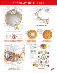
- Page 87: Eyeball
- Page 88: Anterior and Posterior Chambers of Eye
- Page 89: Lens and Supporting Structures
- Page 90: Intrinsic Arteries and Veins of Eye
- Page 91: Vascular Supply of Eye
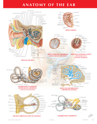
- Page 92: Pathway of Sound Reception
- Page 93: External Ear and Tympanic Cavity
- Page 94: Tympanic Cavity
- Page 95: Bony and Membranous Labyrinths
- Page 96: Bony and Membranous Labyrinths
- Page 97: Orientation of Labyrinth in Skull
- Page 98: Auditory (Pharyngotympanic, Eustachian) Tube
- Page 99: Meninges and Diploic Veins
- Page 100: Meningeal Arteries
- Page 101: Epidural Hematoma
- Page 102: Meninges and Superficial Cerebral Veins
- Page 103: Dural Venous Sinuses
- Page 104: Dural Venous Sinuses
- Page 105: Cerebrum: Lateral Views
- Page 106: Cerebrum: Medial Views
- Page 107: Cerebrum: Inferior View
- Page 108: Ventricles of Brain
- Page 109: Circulation of Cerebrospinal Fluid
- Page 110: Basal Nuclei (Ganglia)
- Page 111: Thalamus
- Page 112: Hippocampus and Fornix
- Page 113: Cerebellum
- Page 114: Brainstem
- Page 115: Fourth Ventricle and Cerebellum
- Page 116: Cranial Nerve Nuclei in Brainstem: Schema
- Page 117: Cranial Nerve Nuclei in Brainstem: Schema
- Page 118: Cranial Nerves (Motor and Sensory Distribution): Schema
- Page 119: Olfactory Nerve (I): Schema
- Page 120: Optic Nerve (II) (Visual Pathway): Schema
- Page 121: Oculomotor (III), Trochlear (IV) and Abducent Nerves (VI): Schema
- Page 122: Trigeminal Nerve (V): Schema
- Page 123: Facial Nerve (VII): Schema
- Page 124: Vestibulocochlear Nerve (VIII): Schema
- Page 125: Glossopharyngeal Nerve (IX): Schema
- Page 126: Vagus Nerve (X): Schema
- Page 127: Accessory Nerve (XI): Schema
- Page 128: Hypoglossal Nerve (XII): Schema
- Page 129: Cervical Plexus: Schema
- Page 130: Autonomic Nerves in Neck
- Page 131: Autonomic Nerves in Head
- Page 132: Ciliary Ganglion: Schema
- Page 133: Pterygopalatine and Submandibular Ganglia: Schema
- Page 134: Otic Ganglion: Schema
- Page 135: Taste Pathways: Schema
- Page 136: Arteries to Brain and Meninges
- Page 137: Veins of the Vertebral Column: Vertebral Veins
- Page 138: Arteries to Brain: Schema
- Page 139: Arteries of Brain: Inferior Views
- Page 140: Cerebral Arterial Circle (Willis)
- Page 141: Arteries of Brain: Frontal View and Section
- Page 142: Arteries of Brain: Lateral and Medial Views
- Page 143: Arteries of Posterior Cranial Fossa
- Page 144: Veins of Posterior Cranial Fossa
- Page 145: Deep Veins of Brain
- Page 146: Subependymal Veins of Brain
- Page 147: Hypothalamus and Hypophysis
- Page 148: Arteries and Veins of Hypothalamus and Hypophysis
- Page 149: Head Scans: Sagittal MR Images
- Page 150: Head Scans: Axial CT Images
- Page 151: Head Scans: Coronal CT Images
- Page 152: Back
- Page 153: Vertebral Column
- Page 154: Thoracic Vertebrae
- Page 155: Lumbar Vertebrae
- Page 156: Lumbar Vertebrae: Radiographs
- Page 157: Sacrum and Coccyx
- Page 158: Vertebral Ligaments: Lumbar Region
- Page 159: Vertebral Ligaments: Lumbosacral Region
- Page 160: Spinal Cord and Ventral Rami In Situ
- Page 161: Relation of Spinal Nerve Roots to Vertebrae
- Page 162: Lumbar Disc Herniation: Clinical Manifestations
- Page 163: Lumbar Puncture and Epidural Anesthesia
- Page 164: Dermatomes
- Page 165: Sympathetic Nervous System: General Topography
- Page 166: Parasympathetic Nervous System: General Topography
- Page 167: Sympathetic Nervous System: Schema
- Page 168: Parasympathetic Nervous System: Schema
- Page 169: Spinal Membranes and Nerve Roots
- Page 170: Spinal Nerve Origin: Cross Sections
- Page 171: Arteries of Spinal Cord: Schema
- Page 172: Arteries of Spinal Cord: Intrinsic Distribution
- Page 173: Veins of Spinal Cord and Vertebral Column
- Page 174: Muscles of Back: Superficial Layers
- Page 175: Muscles of Back: Intermediate Layers
- Page 176: Muscles of Back: Deep Layers
- Page 177: Nerves of Back
- Page 178: Suboccipital Triangle
- Page 179: Lumbar Region of Back: Cross Section
- Page 180: Typical Thoracic Spinal Nerve
- Page 181: Thorax
- Page 182: Mammary Gland
- Page 183: Arteries of Mammary Gland
- Page 184: Lymph Vessels and Nodes of Mammary Gland
- Page 185: Bony Framework of Thorax
- Page 186: Ribs and Sternocostal Joints
- Page 187: Costovertebral Joints
- Page 188: Anterior Thoracic Wall
- Page 189: Anterior Thoracic Wall
- Page 190: Chest Drainage Tube Placement
- Page 191: Anterior Thoracic Wall: Internal View
- Page 192: Intercostal Nerves and Arteries
- Page 193: Phrenic Nerve
- Page 194: Diaphragm: Thoracic Surface
- Page 195: Diaphragm: Abdominal Surface
- Page 196: Topography of Lungs: Anterior View
- Page 197: Topography of Lungs: Posterior View
- Page 198: Lungs In Situ: Anterior View
- Page 199: Lungs: Medial Views
- Page 200: Bronchopulmonary Segments
- Page 201: Bronchopulmonary Segments
- Page 202: Trachea and Major Bronchi
- Page 203: Nomenclature of Bronchi: Schema
- Page 204: Intrapulmonary Airways: Schema
- Page 205: Intrapulmonary Blood Circulation: Schema
- Page 206: Great Vessels of Superior Mediastinum
- Page 207: Bronchial Arteries and Veins
- Page 208: Lymph Vessels and Nodes of Lung
- Page 209: Autonomic Nerves in Thorax
- Page 210: Innervation of Tracheobronchial Tree: Schema
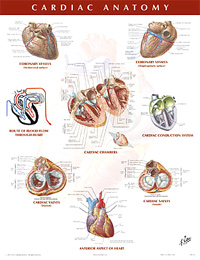
- Page 211: Heart In Situ
- Page 212: Heart: Anterior Exposure
- Page 213: Radiograph of Chest
- Page 214: Heart: Base and Diaphragmatic Surfaces
- Page 215: Pericardial Sac
- Page 216: Coronary Arteries and Cardiac Veins
- Page 217: Coronary Arteries and Cardiac Veins: Variations
- Page 218: Coronary Arteries: Arteriographic Views
- Page 219: Coronary Arteries: Arteriographic Views
- Page 220: Right Atrium and Ventricle
- Page 221: Left Atrium and Ventricle
- Page 222: Valves and Fibrous Skeleton of Heart
- Page 223: Valves and Fibrous Skeleton of Heart
- Page 224: Atria, Ventricles and Interventricular Septum
- Page 225: Conducting System of Heart
- Page 226: Nerves of Heart
- Page 227: Innervation of Heart: Schema
- Page 228: Innervation of Blood Vessels: Schema
- Page 229: Prenatal and Postnatal Circulation
- Page 230: Mediastinum: Right Lateral View
- Page 231: Mediastinum: Left Lateral View
- Page 232: Esophagus In Situ
- Page 233: Topography and Constrictions of Esophagus
- Page 234: Musculature of Esophagus
- Page 235: Pharyngoesophageal Junction
- Page 236: Esophagogastric Junction
- Page 237: Arteries of Esophagus
- Page 238: Veins of Esophagus
- Page 239: Lymph Vessels and Nodes of Esophagus
- Page 240: Nerves of Esophagus
- Page 241: Mediastinum: Cross Section (Superior View)
- Page 242: Chest Scans: Axial CT Images
- Page 243: Cross Section of Thorax at T3 Level
- Page 244: Cross Section of Thorax at T3-4 Disc Level
- Page 245: Cross Section of Thorax at T4-5 Disc Level
- Page 246: Cross Section of Thorax at T7 Level
- Page 247: Abdomen
- Page 248: Bony Framework of Abdomen
- Page 249: Anterior Abdominal Wall: Superficial Dissection
- Page 250: Anterior Abdominal Wall: Intermediate Dissection
- Page 251: Anterior Abdominal Wall: Deep Dissection
- Page 252: Rectus Sheath: Cross Sections
- Page 253: Anterior Abdominal Wall: Internal View
- Page 254: Posterolateral Abdominal Wall
- Page 255: Arteries of Anterior Abdominal Wall
- Page 256: Veins of Anterior Abdominal Wall
- Page 257: Nerves of Anterior Abdominal Wall
- Page 258: Thoracoabdominal Nerves
- Page 259: Inguinal Region: Dissections
- Page 260: Inguinal Canal and Spermatic Cord
- Page 261: Indirect Inguinal Hernia
- Page 262: Femoral Sheath and Inguinal Canal
- Page 263: Posterior Abdominal Wall: Internal View
- Page 264: Arteries of Posterior Abdominal Wall
- Page 265: Veins of Posterior Abdominal Wall
- Page 266: Lymph Vessels and Nodes of Posterior Abdominal Wall
- Page 267: Nerves of Posterior Abdominal Wall
- Page 268: Regions and Planes of Abdomen
- Page 269: Greater Omentum and Abdominal Viscera
- Page 270: Mesenteric Relations of Intestines
- Page 271: Mesenteric Relations of Intestines
- Page 272: Omental Bursa: Stomach Reflected
- Page 273: Omental Bursa: Cross Section
- Page 274: Peritoneum of Posterior Abdominal Wall
- Page 275: Stomach In Situ
- Page 276: Mucosa of Stomach
- Page 277: Musculature of Stomach
- Page 278: Duodenum In Situ
- Page 279: Mucosa and Musculature of Duodenum
- Page 280: Mucosa and Musculature of Small Intestine
- Page 281: Ileocecal Region
- Page 282: Ileocecal Region
- Page 283: (Vermiform) Appendix
- Page 284: Mucosa and Musculature of Large Intestine
- Page 285: Sigmoid Colon: Variations in Position
- Page 286: Topography of Liver
- Page 287: Surfaces and Bed of Liver
- Page 288: Liver In Situ and Variations in Form
- Page 289: Liver Segments and Lobes: Vessel and Duct Distribution
- Page 290: Intrahepatic Vascular and Duct Systems
- Page 291: Liver Structure: Schema
- Page 292: Intrahepatic Biliary System: Schema
- Page 293: Schematic Cross Section of Abdomen at T10
- Page 294: Gallbladder and Extrahepatic Bile Ducts
- Page 295: Junction of (Common) Bile Duct and Duodenum
- Page 296: Variations in Cystic and Hepatic Ducts
- Page 297: Schematic Cross Section of Abdomen at T12
- Page 298: Pancreas In Situ
- Page 299: Spleen
- Page 300: Arteries of Stomach, Liver and Spleen
- Page 301: Arteries of Liver, Pancreas, Duodenum and Spleen
- Page 302: Variations in Celiac Trunk
- Page 303: Celiac Arteriogram
- Page 304: Arteries of Duodenum and Head of Pancreas
- Page 305: Arterial Variations and Collateral Supply of Liver and Gallbladder
- Page 306: Arteries of Small Intestine
- Page 307: Arteries of Large Intestine
- Page 308: Variations in Colic Arteries
- Page 309: Veins of Stomach, Duodenum, Pancreas and Spleen
- Page 310: Veins of Small Intestine
- Page 311: Veins of Large Intestine
- Page 312: Hepatic Portal Vein Tributaries: Portocaval Anastomoses
- Page 313: Variations of Hepatic Portal Vein
- Page 314: Lymph Vessels and Nodes of Stomach
- Page 315: Lymph Vessels and Nodes of Pancreas
- Page 316: Lymph Vessels and Nodes of Small Intestine
- Page 317: Lymph Vessels and Nodes of Large Intestine
- Page 318: Autonomic Nerves and Ganglia of Abdomen
- Page 319: Nerves of Stomach and Duodenum
- Page 320: Nerves of Stomach and Duodenum
- Page 321: Innervation of Stomach and Duodenum: Schema
- Page 322: Nerves of Small Intestine
- Page 323: Nerves of Large Intestine
- Page 324: Innervation of Small and Large Intestines: Schema
- Page 325: Autonomic Reflex Pathways: Schema
- Page 326: Intrinsic Autonomic Plexuses of Intestine: Schema
- Page 327: Innervation of Liver and Biliary Tract: Schema
- Page 328: Innervation of Pancreas: Schema
- Page 329: Kidneys In Situ: Anterior Views
- Page 330: Kidneys In Situ: Posterior Views
- Page 331: Schematic Cross Section of Abdomen at T12
- Page 332: Renal Artery and Vein In Situ
- Page 333: Variations in Renal Artery and Vein
- Page 334: Gross Structure of Kidney
- Page 335: Intrarenal Arteries and Renal Segments
- Page 336: Nephron and Collecting Tubule: Schema
- Page 337: Blood Vessels in Parenchyma of Kidney: Schema
- Page 338: Schematic Cross Section of Abdomen at T12-L1
- Page 339: Schematic Cross Section of Abdomen at L1-2
- Page 340: Ureters
- Page 341: Arteries of Ureters and Urinary Bladder
- Page 342: Renal Fascia
- Page 343: Lymph Vessels and Nodes of Kidneys and Urinary Bladder
- Page 344: Nerves of Kidneys, Ureters and Urinary Bladder
- Page 345: Innervation of Kidneys and Upper Ureters: Schema
- Page 346: Nerves of Suprarenal Glands: Dissection and Schema
- Page 347: Arteries and Veins of Suprarenal Glands In Situ
- Page 348: Abdominal Wall and Viscera: Paramedian (Sagittal) Section
- Page 349: Schematic Cross Section of Abdomen at L3,4
- Page 350: Abdominal Scans: Axial CT Images
- Page 351: Pelvis and Perineum
- Page 352: Bones and Ligaments of Pelvis
- Page 353: Bones and Ligaments of Pelvis
- Page 354: Sex Differences of Pelvis: Measurements
- Page 355: Female and Male Pelvis Radiographs
- Page 356: Pelvic Diaphragm: Female
- Page 357: Pelvic Diaphragm: Female
- Page 358: Pelvic Diaphragm: Male
- Page 359: Pelvic Diaphragm: Male
- Page 360: Pelvic Viscera and Perineum: Female
- Page 361: Pelvic Viscera and Perineum: Male
- Page 362: Pelvic Contents: Female
- Page 363: Pelvic Contents: Male
- Page 364: Endopelvic Fascia and Potential Spaces
- Page 365: Urinary Bladder: Orientation and Supports
- Page 366: Urinary Bladder: Female and Male
- Page 367: Sphincters
- Page 368: Male and Female Cystourethrograms
- Page 369: Pelvic Viscera: Female
- Page 370: Uterus, Vagina and Supporting Structures
- Page 371: Uterus and Adnexa
- Page 372: Pelvic Ligaments
- Page 373: Uterus: Age Changes and Muscle Pattern
- Page 374: Uterus: Variations in Position
- Page 375: Ectopic Pregnancy
- Page 376: Male Pelvis: Bladder-Prostate Junction
- Page 377: Female Perineum and External Genitalia (Pudendum or Vulva)
- Page 378: Female Perineum (Superficial Dissection)
- Page 379: Female Perineum and Deep Perineum
- Page 380: Perineum and External Genitalia: Superficial Dissection
- Page 381: Perineum and External Genitalia (Deeper Dissection)
- Page 382: Penis
- Page 383: Perineal Spaces
- Page 384: Prostate and Seminal Vesicles
- Page 385: Urethra
- Page 386: Descent of Testis
- Page 387: Scrotum and Contents
- Page 388: Homologues of External Genitalia
- Page 389: Homologues of Internal Genitalia
- Page 390: Testis, Epididymis and Ductus Deferens
- Page 391: Rectum In Situ: Female and Male
- Page 392: Ischioanal Fossae
- Page 393: Rectum and Anal Canal
- Page 394: Anorectal Musculature
- Page 395: External Anal Sphincter Muscle: Perineal Views
- Page 396: Actual and Potential Perineopelvic Spaces
- Page 397: Pelvic Scans: Sagittal MR Images
- Page 398: Arteries of Rectum and Anal Canal
- Page 399: Veins of Rectum and Anal Canal
- Page 400: Arteries and Veins of Pelvic Organs: Female
- Page 401: Arteries and Veins of Testis
- Page 402: Arteries and Veins of Pelvis: Female
- Page 403: Arteries and Veins of Pelvis: Male
- Page 404: Arteries and Veins of Perineum and Uterus
- Page 405: Arteries and Veins of Perineum: Male
- Page 406: Lymph Vessels and Nodes of Pelvis and Genitalia: Female
- Page 407: Lymph Vessels and Nodes of Perineum: Female
- Page 408: Lymph Vessels and Nodes of Pelvis and Genitalia: Male
- Page 409: Nerves of External Genitalia: Male
- Page 410: Nerves of Pelvic Viscera: Male
- Page 411: Nerves of Perineum: Male
- Page 412: Nerves of Pelvic Viscera: Female
- Page 413: Nerves of Perineum and External Genitalia: Female
- Page 414: Neuropathways in Parturition
- Page 415: Innervation of Female Reproductive Organs: Schema
- Page 416: Innervation of Male Reproductive Organs: Schema
- Page 417: Innervation of Urinary Bladder and Lower Ureter: Schema
- Page 418: Upper Limb
- Page 419: Clavicle and Sternoclavicular Joint
- Page 420: Humerus and Scapula: Anterior Views
- Page 421: Humerus and Scapula: Posterior Views
- Page 422: Shoulder: Anteroposterior Radiograph
- Page 423: Shoulder (Glenohumeral) Joint
- Page 424: Muscles of Shoulder
- Page 425: Muscles of Rotator Cuff
- Page 426: Scapulohumeral Dissection
- Page 427: Axillary Artery and Anastomoses Around Scapula
- Page 428: Pectoral, Clavipectoral and Axillary Fasciae
- Page 429: Axilla (Dissection): Anterior View
- Page 430: Brachial Plexus: Schema
- Page 431: Muscles of Arm: Anterior Views
- Page 432: Muscles of Arm: Posterior View
- Page 433: Brachial Artery In Situ
- Page 434: Brachial Artery and Anastomoses Around Elbow
- Page 435: Arm: Serial Cross Sections
- Page 436: Bones of Elbow
- Page 437: Elbow: Radiographs
- Page 438: Ligaments of Elbow
- Page 439: Bones of Forearm
- Page 440: Individual Muscles of Forearm: Rotators of Radius
- Page 441: Individual Muscles of Forearm: Extensors of Wrist and Digits
- Page 442: Individual Muscles of Forearm: Flexors of Wrist
- Page 443: Individual Muscles of Forearm: Flexors of Digits
- Page 444: Muscles of Forearm (Superficial Layer): Posterior View
- Page 445: Muscles of Forearm (Deep Layer): Posterior View
- Page 446: Muscles of Forearm (Superficial Layer): Anterior View
- Page 447: Muscles of Forearm (Intermediate Layer): Anterior View
- Page 448: Muscles of Forearm (Deep Layer): Anterior View
- Page 449: Forearm: Serial Cross Sections
- Page 450: Attachments of Muscles of Forearm: Anterior View
- Page 451: Attachments of Muscles of Forearm: Posterior View
- Page 452: Carpal Bones
- Page 453: Movements of Wrist
- Page 454: Ligaments of Wrist
- Page 455: Ligaments of Wrist
- Page 456: Bones of Wrist and Hand
- Page 457: Wrist and Hand: Anteroposterior Radiograph
- Page 458: Metacarpophalangeal and Interphalangeal Ligaments
- Page 459: Wrist and Hand: Superficial Palmar Dissection
- Page 460: Wrist and Hand: Deeper Palmar Dissections
- Page 461: Flexor Tendons, Arteries and Nerves at Wrist
- Page 462: Bursae, Spaces and Tendon Sheaths of Hand
- Page 463: Lumbrical Muscles, Bursae, Spaces and Sheaths: Schema
- Page 464: Flexor and Extensor Tendons in Fingers
- Page 465: Intrinsic Muscles of Hand
- Page 466: Arteries and Nerves of Hand: Palmar Views
- Page 467: Wrist and Hand: Superficial Radial Dissection
- Page 468: Wrist and Hand: Superficial Dorsal Dissection
- Page 469: Wrist and Hand: Deep Dorsal Dissection
- Page 470: Extensor Tendons at Wrist
- Page 471: Fingers
- Page 472: Cutaneous Innervation of Wrist and Hand
- Page 473: Arteries and Nerves of Upper Limb
- Page 474: Musculocutaneous Nerve
- Page 475: Median Nerve
- Page 476: Ulnar Nerve
- Page 477: Radial Nerve in Arm and Nerves of Posterior Shoulder
- Page 478: Radial Nerve in Forearm
- Page 479: Cutaneous Nerves and Superficial Veins of Shoulder and Arm
- Page 480: Cutaneous Nerves and Superficial Veins of Forearm
- Page 481: Cutaneous Innervation of Upper Limb
- Page 482: Dermatomes of Upper Limb
- Page 483: Lymph Vessels and Nodes of Upper Limb
- Page 484: CT Angiograms of Arteries of the Upper limb
- Page 485: Lower Limb
- Page 486: Hip (Coxal) Bone
- Page 487: Hip Joint
- Page 488: Hip Joint: Anteroposterior Radiograph
- Page 489: Femur
- Page 490: Bony Attachments of Muscles of Hip and Thigh: Anterior View
- Page 491: Bony Attachments of Muscles of Hip and Thigh: Posterior View
- Page 492: Muscles of Thigh: Anterior Views
- Page 493: Muscles of Thigh: Anterior Views
- Page 494: Muscles of Hip and Thigh: Lateral View
- Page 495: Muscles of Hip and Thigh: Posterior Views
- Page 496: Psoas and Iliacus Muscles
- Page 497: Lumbosacral and Coccygeal Plexuses
- Page 498: Lumbar Plexus
- Page 499: Sacral and Coccygeal Plexuses
- Page 500: Arteries and Nerves of Thigh: Anterior Views
- Page 501: Arteries and Nerves of Thigh: Anterior Views
- Page 502: Arteries and Nerves of Thigh: Posterior View
- Page 503: Nerves of Hip and Buttock
- Page 504: Arteries of Femoral Head and Neck
- Page 505: Thigh: Serial Cross Sections
- Page 506: Knee: Lateral and Medial Views
- Page 507: Knee: Anterior Views
- Page 508: Knee: Interior
- Page 509: Knee: Cruciate and Collateral Ligaments
- Page 510: Knee: Anteroposterior Radiograph
- Page 511: Right Knee: Posterior and Sagittal Views
- Page 512: Arteries of Thigh and Knee: Schema
- Page 513: Tibia and Fibula
- Page 514: Tibia and Fibula
- Page 515: Attachments of Muscles of Leg
- Page 516: Muscles of Leg (Superficial Dissection): Posterior View
- Page 517: Muscles of Leg (Intermediate Dissection): Posterior View
- Page 518: Muscles of Leg (Deep Dissection): Posterior View
- Page 519: Muscles of Leg (Superficial Dissection): Anterior View
- Page 520: Muscles of Leg (Deep Dissection): Anterior View
- Page 521: Muscles of Leg: Lateral View
- Page 522: Leg: Cross Sections and Fascial Compartments
- Page 523: Bones of Foot
- Page 524: Bones of Foot
- Page 525: Calcaneus
- Page 526: Ankle: Radiographs
- Page 527: Ligaments and Tendons of Ankle
- Page 528: Ligaments and Tendons of Foot: Plantar View
- Page 529: Tendon Sheaths of Ankle
- Page 530: Muscles of Dorsum of Foot: Superficial Dissection
- Page 531: Dorsum of Foot: Deep Dissection
- Page 532: Sole of Foot: Superficial Dissection
- Page 533: Muscles of Sole of Foot: First Layer
- Page 534: Muscles of Sole of Foot: Second Layer
- Page 535: Muscles of Sole of Foot: Third Layer
- Page 536: Interosseous Muscles and Deep Arteries of Foot
- Page 537: Interosseous Muscles of Foot
- Page 538: Femoral Nerve and Lateral Cutaneous Nerve of Thigh
- Page 539: Obturator Nerve
- Page 540: Sciatic Nerve and Posterior Cutaneous Nerve of Thigh
- Page 541: Tibial Nerve
- Page 542: Common Fibular (Peroneal) Nerve
- Page 543: Dermatomes of Lower Limb
- Page 544: Superficial Nerves and Veins of Lower Limb: Anterior View
- Page 545: Superficial Nerves and Veins of Lower Limb: Posterior View
- Page 546: Lymph Vessels and Nodes of Lower Limb
- Page 547: CT Angiograms of Arteries of the Lower Limb