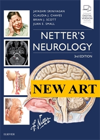
New Art: Netter's Neurology 3E
Author: Jayashri Srinivasan
ISBN: 9780323554763
- Page 40: Hounsfield Units (HU) and brain imaging.
- Page 41: Computed Tomography (CT) Reconstruction Algorithm and CT Window
- Page 41.1: Computed Tomography (CT) Window Level and Width
- Page 42: Clinical Example of the Value of Windowing
- Page 42.1: Computed Tomography Postprocessing
- Page 43: Computed Tomography Angiography (CTA), Computed Tomography Venography (CTV), and Computed Tomography Perfusion (CTP)
- Page 43.1: Substances and Tissues Have Characteristic T1 and T2 Signal Intensities
- Page 44: Different magnetic resonance imaging sequences accentuate the differences that tissue injury or disease creates within the central nervous system
- Page 45: Magnetic Resonance Spectroscopy (MRS)
- Page 45.1: Functional Magnetic Resonance Imaging (MRI)
- Page 46: Vascular Ultrasound of the Carotid Bifurcations
- Page 47: X-ray images are useful due to their high spatial resolution and lack of CT and MRI related artifacts.
- Page 47.1: Digital Subtraction Angiography
- Page 48: Nuclear Medicine
- Page 134: Noncontrast Head Computed Tomography
- Page 136: Brain in a Box Model
- Page 136.1: Intracranial Pressure/Herniation
- Page 137: Malignant Middle Cerebral Artery Ischemic Stroke
- Page 137.1: Hemorrhagic Conversion of Right Middle Cerebral Artery Ischemic Stroke After Tissue Plasminogen Activator
- Page 146: Quantitative Electroencephalography Time Lapsed Over 45 Minutes
- Page 148: Intracranial Pressure (ICP) Waveform
- Page 153: Initial Management of Coma and Severe Head Injuries
- Page 153.1: Wernicke Encephalopathy With MRI T2 Fluid-Attenuated Inversion Recovery (FLAIR) Changes Involving Medial Thalamus (1), Mammillary Bodies (2), and Periaqueductal Gray (3)
- Page 154: Triphasic Waves on EEG as Seen in Metabolic and Hepatic Coma
- Page 173: Cardiac Embolism
- Page 174: Lacunar Infarction
- Page 174: Arterial Dissection
- Page 185: Mechanical Thrombectomy of Large Vessel Occlusion
- Page 191: Images of a Typical Hypertensive Putaminal Intracerebral Hemorrhage
- Page 191.1: Hypertensive Intracerebral Hemorrhage: Pathogenesis
- Page 192: Typical CT (A) and susceptibility-weighted MR imaging (B and C) of intracerebral hemorrhage (ICH) due to probable cerebral amyloid angiopathy. There is a right frontal cortico-subcortical hematoma with overlying subarachnoid hemorrhage (A and B), as well as numerous, predominantly cortico-subcortical microhemorrhages (hypointense dots) in both the region of the ICH (B) and the posterior regions of the cerebral hemispheres (C)
- Page 193: Typical CT (panel A) and susceptibility-weighted MR imaging (panels B and C) of ICH due to probable cerebral amyloid angiopathy
- Page 194: Pathophysiology of Brain Injury Caused by Intracerebral Hemorrhage
- Page 196: Clinical Presentation of Deep Supratentorial Intracerebral Hemorrhage by Anatomic Location
- Page 199: Structural complications from thalamic (panels A and B) and cerebellar (panels D and E) intracerebral hemorrhages on CT
- Page 200: Brainstem Intracerebral Hemorrhage
- Page 208: Meninges and Their Relationship to the Brain Parenchyma. Top: Layers of the scalp, bone, and meninges in relation to the brain parenchyma. Botton: Blown up image of the meninges in relation to the brain. Green: pia mater, Pink: subarachnoid space
- Page 209: Perimesencephalic Subarachnoid Hemorrhage
- Page 212: A, Reversible Cerebral Vasoconstriction Syndrome Subarachnoid Hemorrhage. Axial noncontrast head CT with hyperdensity in the left frontal sulci consistent with cortical subarachnoid hemorrhage. B, Reversible Cerebral Vasoconstriction Syndrome - Aniography. Left internal carotid artery injection with demonstration of multifocal narrowing of multiple distal branches in the left middle cerebral artery (red arrows) territory consistent with reversible cerebral vasoconstriction syndrome
- Page 215: Interventional Radiologic Repair of Berry Aneurysm
- Page 218: Changes Associated With Subarachnoid Hemorrhage
- Page 270: Pain Pathways
- Page 271: Physiology of Nociceptor Activation
- Page 352: Clinical Signs of Parkinson Disease
- Page 354: Nonmotor Symptoms of Parkinson Disease
- Page 407: CIS Case
- Page 407.1: RIS Case
- Page 408: RRMS Case
- Page 409: SPMS Case
- Page 411: Visual Evoked Response (VER)
- Page 418: (A) Sagittal T2 MRI of the thoracic spinal cord showing extensive abnormal T2 hyperintense signal in the lower cervical and upper thoracic cord with more than three segments involved, characteristic of NMO. (B) Sagittal T1 Fat Sat postcontrast image demonstrating peripheral intramedullary enhancement from T2 through T4
- Page 420: (A,B) MRI of the brain showing prominent T2 hyperintensity involving the left frontal and parietal lobes with extension to the internal capsule, midbrain and pons. (C,D) Postgadolinium enhanced T1 showing peripheral enhancement. (E,F) MRI of the brain performed 1 year after the initial presentation showing significant improvement of the T2 hyperintensities with residual abnormalities in the left peri-rolandic area and underlying white matter
- Page 421: Acute Disseminated Encephalomyelitis
- Page 458: Lyme Disease
- Page 529: Axial, Gadolinium Contrast–Enhanced T1-Weighted Magnetic Resonance Image Shows 6-mm Enhancing Metastasis in Left Cerebellar Hemisphere
- Page 530: Axial, Gadolinium Contrast–Enhanced T1-Weighted Magnetic Resonance Image of Lower Cervical Spine Shows Enhancing Anterior and Posterior Nerve Roots
- Page 530.1: Positron Emission Tomography–Computed Tomography Scan Shows Intense Fluorodeoxyglucose Uptake in Bilateral Mediastinal, Hilar, and Subcarinal Lymph Nodes
- Page 532: Paraneoplastic Disorder of Peripheral Nervous System
- Page 560: Acute Cervical Spinal Demyelinating Plaque in a Patient With Multiple Sclerosis
- Page 560.1: Acute Myelitis in a Patient With Neuromyelitis Optica Spectrum Disorder
- Page 564: Subacute Combined Degeneration
- Page 565: Metastatic Malignancies
- Page 565.1: HTLV-1 Myelopathy
- Page 566: Extramedullary Intradural Spinal Tumors
- Page 572: Metastatic Malignances
- Page 684: Acetylcholine Receptor and Neuromuscular Junction
- Page 685: Immunopathology of Myasthenia Gravis
- Page 686: Thymoma
