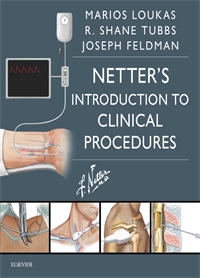
Clinical Procedures - Loukas 1st Edition
Author: Marios Loukas
ISBN: 9780323370554
- Page 10: Bony framework of thorax. Intercostal nerves and arteries
- Page 11: Spinal cord and nerves of anterior abdominal wall
- Page 12: Dermatomes above the waist
- Page 13: Intercostal Nerve Block
- Page 16: Airway anatomy
- Page 17: Larynx and tongue
- Page 19: Bag-valve-mask ventilation
- Page 22: Contents of the thorax: mediastinum
- Page 23: Mediastinum
- Page 24: Imaging of mediastinum
- Page 27: Thoracostomy
- Page 30: Topography of lungs: posterior view
- Page 31: Intercostal nerves and arteries
- Page 33: Ultrasonography for thoracentesis
- Page 35: Thoracentesis
- Page 38: Cricoid cartilage
- Page 39: Thyroid gland and suprahyoid and infrahyoid muscles
- Page 41: Open tracheotomy
- Page 46: Heart: anterior exposure
- Page 47: Right atrium and ventricle
- Page 48: Conducting system of the heart
- Page 49: Cardiac depolarization and repolarization
- Page 51: Transvenous pacemaker placement
- Page 62: Anterior abdominal wall: intermediate dissection
- Page 63: Rectus sheath: cross section
- Page 65: Diagnostic peritoneal lavage
- Page 68: Regions and Planes of Abdomen
- Page 69: Subxypoid cardiac view
- Page 70: FAST: Right Flank Examination
- Page 71: Left upper quadrant view
- Page 72: E-FAST: Suprapubic Examination
- Page 73: E-FAST Thoracic Examination
- Page 76: Leg: fascial compartments
- Page 77: Leg: cross section
- Page 78: Forearm: Serial Cross Sections
- Page 80: Leg Fasciotomy: Double Incision
- Page 81: Forearm Fasciotomy
- Page 89: Pericardium and heart in middle mediastinum just posterior to the sternum. Note the relationship between the sternum, diaphragm, and heart.
- Page 90: A, Pericardial sac surrounds the heart and is innervated by the phrenic nerve. B, Pericardium is removed and the anterior portion of the heart exposed.
- Page 91: Parasternal pericardiocentesis technique
- Page 93: Pericardiocentesis
- Page 105: Arrangement of tendons, vessels, and nerves at the wrist
- Page 106: Wrist and hand: superficial radial dissection
- Page 107: Arteries of upper limb
- Page 109: Peripheral arterial line placement
- Page 119: Cardiovascular System: Major Veins
- Page 120: Cutaenous Nerves and Superficial Veins of Leg
- Page 121: Cutaneous nerves and superficial veins of the forearm
- Page 123: Venous cutdown: distal great saphenous vein
- Page 124: Venous cutdown: forearm veins
- Page 129: Dislocated Hip Reduction: Allis Maneuver
- Page 130: Dislocated Hip Reduction: Stimson Maneuver
- Page 140: Anatomy of the Knee: Medial and Anterior Views
- Page 141: Dislocated Knee Reduction
- Page 145: Shoulder (Glenohumeral Joint)
- Page 146: Axilla and Muscles of Rotator Cuff
- Page 147: Ultrasound of normal (A) and anteriorly dislocated (B and C) shoulder joint
- Page 148: Dislocated Shoulder Joint Reduction : Scapular Manipulation
- Page 149: Dislocated Shoulder Joint Reduction : Lateral Rotation Maneuver
- Page 152: Occipital nerve anatomy
- Page 153: Cutaneous nerves of the head and neck
- Page 155: Occipital Nerve Block
- Page 157: Hand, foot, and finger dissections
- Page 158: Finger Web Space Block
- Page 159: Three-sided Toe Block
- Page 160: Wing Block
- Page 163: Oral Cavity
- Page 164: Teeth and Mandible
- Page 165: Superior Alveolar Nerve Block
- Page 166: Inferior Alveolar Nerve Block
- Page 169: Anterior abdominal wall: deep dissection
- Page 170: Regions and planes of the abdomen
- Page 171: Ascites
- Page 172: Imaging of abdominal paracentesis
- Page 173: Abdominal Paracentesis
- Page 175: Right auricle (pinna)
- Page 176: Tympanic membrane
- Page 177: Tympanic cavity
- Page 179: Incision and drainage for auricular hematoma
- Page 182: Anatomy of the Ear
- Page 183: Coronal oblique section of external acoustic meatus and middle ear (tympanic cavity)
- Page 184: Cerumen removal: ear irrigation
- Page 185: Cerumen removal: blunt ear curettes
- Page 187: Lateral Wall of Nasal Cavity
- Page 188: Anatomy of the Ear
- Page 189: Direct Removal of Foreign Body in Nose
- Page 193: Superficial Arteries and Veins of Face and Scalp
- Page 194: Meninges and Superficial Cerebral Veins
- Page 195: Hematomas
- Page 196: General Features of Hemorrhage
- Page 201: Bones of Elbow
- Page 202: Ligaments of Elbow
- Page 203: Ultrasound imaging of the elbow joint.�
- Page 204: Ultrasound imaging of normal elbow joint and elbow joint effusion
- Page 205: Elbow Joint Aspiration
- Page 209: Normal Structure and Function of the Nail Unit
- Page 211: Ingrown toenail removal
- Page 214: Anatomy of the Knee
- Page 215: Septic Bursitis and Arthritis
- Page 217: Knee Joint Aspiration
- Page 220: Anatomy of vertebral column
- Page 221: Imaging of the Spine
- Page 223: Lumbar Puncture procedure
- Page 226: Lateral Wall of Nasal Cavity and Pharynx
- Page 227: Contraindication: Midface Fractures
- Page 229: Nasogastric Tube Placement
- Page 232: Anatomy of Fingers
- Page 233: Cross-sectional anatomy and arteries and nerves of the finger
- Page 235: Paronychia incision and drainage
- Page 238: Abscesses
- Page 239: Ultrasound imaging of abscesses
- Page 241: Skin Abscess Incision and Drainage
- Page 243: Shoulder (Glenohumeral Joint)
- Page 244: Shoulder: anterior, superior, and coronal views
- Page 246: Shoulder joint aspiration: anterior approach
- Page 247: Shoulder joint aspiration: posterior approach
