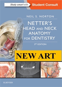
Flash Cards - Advanced Head and Neck, Norton 1E
Author: Neil S. Norton
ISBN: 9781416046318
- Page 1.01: Embyrological Development
- Page 1.02: Pharyngeal Arches
- Page 1.03: Cartilage Derivatives of Pharyngeal Arches
- Page 1.04: Pharyngeal Pouches
- Page 1.05: Development of the Skull
- Page 1.06: Development of the Face
- Page 1.07: Development of the Palate
- Page 1.08: Development of the Tongue
- Page 1.09: Development of the Thyroid
- Page 1.1: Ectopic Thyroid (Pharyngeal Pouch Abnormalities)
- Page 1.11: Cleft Lip and Palate
- Page 2.01: Articulations
- Page 2.02: Articulations
- Page 2.03: Frontal Bone
- Page 2.04: Parietal Bone
- Page 2.05: Occipital Bone
- Page 2.06: Temporal Bone
- Page 2.07: Sphenoid Bone
- Page 2.08: Lacrimal and Nasal Bones
- Page 2.09: Zygomatic Bones
- Page 2.1: Ethmoid Bone
- Page 2.11: Vomer
- Page 2.12: Inferior Nasal Conchae and Palatine Bones
- Page 2.13: Maxilla
- Page 2.14: Mandible
- Page 2.15: Mandible
- Page 2.16: Foramina and Fissures from the Superior View
- Page 2.17: Foramina and Fissures from the Inferior View
- Page 2.18: Foramina and Fissures from the Anterior View
- Page 2.19: Cervical Vertebrae
- Page 2.2: Major External Ligaments
- Page 2.21: Major Internal Ligaments
- Page 2.22: Zygomatic Bones
- Page 2.23: Le Fort Fractures
- Page 2.24: Cervical Fractures
- Page 3.01: Nervous Tissue: Neurons
- Page 3.02: Nervous Tissue: Neuroglia
- Page 3.03: Central Nervous System
- Page 3.04: Peripheral Nervous System
- Page 3.05: Autonomic Nervous System
- Page 3.06: Parasympathetics: Eye
- Page 3.07: Sympathetics: Eye
- Page 3.08: Parasympathetics: Palatine, Pharyngeal, Lacrimal Glands, and Nasal Cavity
- Page 3.09: Sympathetics: Palatine, Pharyngeal, Lacrimal Glands, and Nasal Cavity
- Page 3.1: Parasympathetics: Submandibular and Sublingual Glands
- Page 3.11: Sympathetics: Submandibular and Sublingual Glands
- Page 3.12: Parasympathetics: Parotid Gland
- Page 3.13: Sympathetics: Parotid Gland
- Page 3.14: Cranial Nerves
- Page 4.01: Neck
- Page 4.02: Anterior Triangle
- Page 4.03: Submandibular Triangle
- Page 4.04: Submandibular Triangle: Contents
- Page 4.05: Carotid Triangle
- Page 4.06: Carotid Triangle: Contents
- Page 4.07: Muscular Triangle
- Page 4.08: Muscular Triangle: Contents
- Page 4.09: Submental Triangle
- Page 4.1: Posterior Triangle
- Page 4.11: Posterior Triangle: Contents
- Page 4.12: Suboccipital Triangle
- Page 4.13: Major Visceral Structures
- Page 4.14: Root of the Neck
- Page 4.15: Muscles: Triangles of the Neck
- Page 4.16: Suprahyoid Muscles
- Page 4.17: Infrahyoid Muscles
- Page 4.18: Prevertebral Muscles
- Page 4.19: Suboccipital Muscles
- Page 4.2: Arterial Supply: Subclavian
- Page 4.21: Arterial Supply: Common Carotid
- Page 4.22: Major Venous Drainage
- Page 4.23: Sensory Innervation
- Page 4.24: Cervical Plexus
- Page 4.25: Pharynx
- Page 4.26: Constrictor Muscles of the Pharynx
- Page 4.27: Longitudinal Muscles of the Pharynx
- Page 4.28: Nasopharynx
- Page 4.29: Oropharynx
- Page 4.3: Laryngopharynx
- Page 4.31: Potential Apertures in the Pharyngeal Wall
- Page 4.32: Arterial Supply of the Pharynx
- Page 4.33: Venous Supply of the Pharynx
- Page 4.34: Nerve Supply of the Pharynx
- Page 4.35: Larynx
- Page 4.36: Cartilages of the Larynx
- Page 4.37: Thyroid Cartilage
- Page 4.38: Cricoid Cartilage
- Page 4.39: Arytenoid Cartilage
- Page 4.4: Major Ligaments of the Larynx
- Page 4.41: Muscles of the Larynx
- Page 4.42: Muscles of the Larynx
- Page 4.43: Muscle Actions of the Larynx
- Page 4.44: Arterial Supply to the Larynx
- Page 4.45: Vagus Nerves: Branches
- Page 4.46: Emergency Airway: Cricothyrotomy
- Page 4.47: Laryngitis
- Page 4.48: Fascia of the Neck
- Page 4.49: Fascial Spaces of the Neck
- Page 4.5: Major Fascial Spaces of the Neck
- Page 4.51: Lateral Pharyngeal Space
- Page 5.01: Face
- Page 5.02: Scalp
- Page 5.03: Arteries of the Scalp
- Page 5.04: Sensory Nerves of the Scalp
- Page 5.05: Muscles of Facial Expression
- Page 5.06: Muscles of Facial Expression
- Page 5.07: Muscles of Facial Expression
- Page 5.08: Muscles of Facial Expression
- Page 5.09: Muscles of Facial Expression
- Page 5.1: Muscles of Facial Expression
- Page 5.11: Muscles of Facial Expression
- Page 5.12: Arterial Supply to the Face
- Page 5.13: Arterial Branches of the Face
- Page 5.14: Arterial Branches of the Face: Maxillary Derivatives
- Page 5.15: Arterial Branches of the Face: Ophthalmic Derivatives
- Page 5.16: Venous Drainage to the Face
- Page 5.17: Venous Drainage to the Face
- Page 5.18: Sensory Innervation to the Face
- Page 5.19: Sensory Innervation to the Face: Ophthalmic Division
- Page 5.2: Sensory Innervation to the Face: Maxillary Division
- Page 5.21: Sensory Innervation to the Face: Mandibular Division
- Page 5.22: Sensory Innervation to the Face: Cervical Plexus
- Page 5.23: Facial Nerve
- Page 5.24: Trigeminal Neuralgia
- Page 5.25: Cavernous Sinus Syndrome
- Page 5.26: Recess of the Parotid Fossa
- Page 5.27: Vascular Supply: Parotid Fossa
- Page 5.28: Major Structures of the Parotid Fossa
- Page 5.29: Sensory Nerves of the Parotid Gland
- Page 5.3: Frey's Syndrome
- Page 5.31: Bell's Palsy
- Page 5.32: Fistulae and Sialoceles
- Page 6.01: Borders and Structures: Temporal Fossa
- Page 6.02: Borders and Structures: Infratemporal Fossa
- Page 6.03: Contents of the Infratemporal Fossa
- Page 6.04: Arteries of the Temporal Fossa
- Page 6.05: Veins of the Temporal Fossa
- Page 6.06: Sensory Nerves of the Temporal Fossa
- Page 6.07: Maxillary Artery
- Page 6.08: First Part of the Maxillary Artery
- Page 6.09: Second Part of the Maxillary Artery
- Page 6.1: Veins of the Infratemporal Fossa
- Page 6.11: Motor Branches of the Mandibular Nerve
- Page 6.12: Sensory Branches of the Mandibular Nerve
- Page 6.13: Autonomic and Taste Nerves in the Infratemporal Fossa
- Page 6.14: Muscles of Mastication
- Page 6.15: Muscles of Mastication
- Page 6.16: Muscles of Mastication
- Page 6.17: Muscles of Mastication
- Page 6.18: Arteries of the Muscles of Mastication
- Page 6.19: Nerves of the Muscles of Mastication
- Page 6.2: Compartments of the Temporomandibular Joint
- Page 6.21: Capsule and Collateral Ligaments of the Temporomandibular Joint
- Page 6.22: Extrinsic Ligaments of the Temporomandibular Joint
- Page 6.23: Arteries of the Temporomandibular Joint
- Page 6.24: Veins of the Temporomandibular Joint
- Page 6.25: Sensory Nerves of the Temporomandibular Joint
- Page 6.26: Dislocation of the Mandible
- Page 6.27: Ankylosis
- Page 6.28: Pterygopalatine Fossa
- Page 6.29: Nerves of the Pterygopalatine Fossa
- Page 6.3: Nerves of the Pterygopalatine Ganglion
- Page 6.31: Arteries of the Pterygopalatine Fossa
- Page 7.01: Nose
- Page 7.02: Vascular Supply of the Nose
- Page 7.03: Vascular Supply of the Nose
- Page 7.04: Nerve Supply of the Nose
- Page 7.05: Nasal Cavity
- Page 7.06: Boundaries and Relatinships of the Nasal Cavity
- Page 7.07: Bones of the Lateral Nasal Wall
- Page 7.08: Concha of the Nasal Cavity
- Page 7.09: Arterial Supply of the Nasal Cavity
- Page 7.1: Sensory Innervation of the Nasal Cavity
- Page 7.11: Sensory Innervation of the Nasal Cavity
- Page 7.12: Features of the Paranasal Sinuses
- Page 7.13: Vascular Supply of the Frontal Sinuses
- Page 7.14: Nerve Supply of the Frontal Sinuses
- Page 7.15: Vascular Supply of the Ethmoid Sinuses
- Page 7.16: Nerve Supply of the Ethmoid Sinuses
- Page 7.17: Vascular Supply of the Maxillary Sinuses
- Page 7.18: Nerve Supply of the Maxillary Sinuses
- Page 7.19: Vascular Supply of the Sphenoid Sinuses
- Page 7.2: Nerve Supply of the Spenoid Sinuses
- Page 7.21: Sinusitis
- Page 8.01: Oral Cavity
- Page 8.02: Anatomy of the Lips and Cheeks
- Page 8.03: Vascular Supply of the Lips
- Page 8.04: Nerve Supply of the Lips
- Page 8.05: Boundaries of the Oral Cavity
- Page 8.06: Hard Palate
- Page 8.07: Soft Palate
- Page 8.08: Muscles of the Soft Palate
- Page 8.09: Muscles of the Soft Palate
- Page 8.1: Cheek
- Page 8.11: Floor of the Oral Cavity
- Page 8.12: Teeth
- Page 8.13: Arterial Supply of the Oral Cavity
- Page 8.14: Arterial Supply of the Oral Cavity
- Page 8.15: Arterial Supply of the Teeth
- Page 8.16: Salivary Glands
- Page 8.17: Tongue
- Page 8.18: Dorsal Surface of the Tongue
- Page 8.19: Ventral Surface of the Tongue
- Page 8.2: Extrinsic Muscles of the Tongue
- Page 8.21: Arterial Supply of the Tongue
- Page 8.22: Lesions Involving they Hypoglossal Nerve
- Page 9.01: Ear
- Page 9.02: External Ear
- Page 9.03: External Ear
- Page 9.04: Middle Ear
- Page 9.05: Middle Ear
- Page 9.06: Ear Ossicles
- Page 9.07: Ear Ossicles
- Page 9.08: Inner Ear
- Page 9.09: Inner Ear
- Page 9.1: Cochlear Duct
- Page 9.11: Muscles of the Ear
- Page 9.12: Vascular Supply of the Middle Ear
- Page 9.13: Acute Otitis Externa
- Page 9.14: Acute Otitiis Media
- Page 9.15: Mastoiditis
- Page 10.01: Osteology of the Orbit
- Page 10.02: Openings in the Orbit
- Page 10.03: Eye
- Page 10.04: Eye: Components
- Page 10.05: Eye: Components
- Page 10.06: Muscles of the Eye
- Page 10.07: Muscles of the Eye
- Page 10.08: Nerves of the Eye
- Page 10.09: Ophthalmic Sensory Innervation to the Orbit
- Page 10.1: Ophthalmic Sensory Innervation to the Orbit
- Page 10.11: Maxillary Sensory Innervation to the Orbit
- Page 10.12: Motor Innervation to the Orbit
- Page 10.13: Vascular Suppy to the Orbit (Ophthalmic Derivatives)
- Page 10.14: Vascular Suppy to the Orbit (Ophthalmic Derivatives)
- Page 10.15: Vascular Suppy to the Orbit (Maxillary Derivatives)
- Page 10.16: Lacrimal Apparatus
- Page 10.17: Glaucoma
- Page 11.01: CT Image: Axial View
- Page 11.02: CT Image: Coronal View
- Page 11.03: CT Image: Sagittal View
- Page 11.04: CT Image: Coronal View
- Page 11.05: CT Image: Coronal View
- Page 11.06: CT Image: Coronal View
- Page 11.07: CT Image: Axial View
- Page 11.08: Lateral Radiograph of the Skull
- Page 11.09: Lateral Radiograph of the Skull
- Page 11.1: CT Image: Sagittal Section