The Netter Presenter: Human Anatomy Collection
This product is no longer available, but individual images or image sets may be purchased
ISBN: 9781929007221

- Page 1.001: Head and Neck
- Page 1.002: Skull: Anterior View
- Page 1.003: Skull: Anterior View - Right Orbit: Frontal and Slightly Lateral View
- Page 1.004: Skull: Anteroposterior Radiograph
- Page 1.005: Skull: Lateral View
- Page 1.008: Skull: Midsagittal Section
- Page 1.009: Skull: Nasal Conchae Exposed - Sagittal Section
- Page 1.01: Calvaria: Superior View
- Page 1.011: Calvaria: Inferior View
- Page 1.012: Cranial Base: Inferior View
- Page 1.013: Bones of Cranial Base: Superior View
- Page 1.014: Foramina of Cranial Base: Superior View
- Page 1.015: Skull of Newborn: Lateral View
- Page 1.016: Skull of Newborn: Superior View
- Page 1.017: Bony Framework of Head and Neck: Lateral View
- Page 1.018: Bony Framework of Head and Neck: Mandible Removed - Lateral View
- Page 1.019: Mandible: Anterolateral Superior View
- Page 1.02: Mandible: Left Posterior View
- Page 1.021: Temporomandibular Joint: Lateral and Medial Views
- Page 1.022: Temporomandibular Joint: Joint Action
- Page 1.023: Cervical Vertebrae: Atlas and Axis
- Page 1.024: Cervical Vertebrae (C1-4) Assembled: Posterosuperior View
- Page 1.025: Cervical Vertebrae: Radiograph of Atlantoaxial Joint
- Page 1.026: Cervical Vertebrae (C4 and C7): Superior Views
- Page 1.027: Cervical Vertebrae (C2-T1) - Assembled: Right Lateral View
- Page 1.028: Cervical Vertebrae: Lateral Radiograph
- Page 1.029: External Craniocervical Ligaments: Anterior and Posterior Views
- Page 1.03: External Craniocervical Ligaments: Right Lateral View
- Page 1.031: Internal Craniocervical Ligaments
- Page 1.032: Internal Craniocervical Ligaments
- Page 1.033: Superficial Arteries and Veins of Face and Scalp
- Page 1.034: Superficial Face: Sources of Arterial Supply
- Page 1.035: Cutaneous Nerves of Head and Neck
- Page 1.036: Dermatomes of Head and Neck
- Page 1.037: Facial Nerve Branches and Parotid Gland In Situ
- Page 1.038: Facial Nerve Branches and Parotid Gland Sectioned
- Page 1.039: Muscles of Facial Expression: Lateral View
- Page 1.04: Muscles of Neck: Lateral View
- Page 1.041: Muscles of Neck: Anterior View
- Page 1.042: Infrahyoid and Suprahyoid Muscles
- Page 1.043: Infrahyoid and Suprahyoid Muscles and Their Action: Schema
- Page 1.044: Scalene and Prevertebral Muscles
- Page 1.045: Superficial Veins and Cutaneous Nerves of Neck
- Page 1.046: Cervical Plexus In Situ
- Page 1.047: Cervical Plexus: Schema
- Page 1.048: Subclavian Artery: Right Anterior Dissection
- Page 1.049: Subclavian Artery: Right Lateral Schematic View
- Page 1.05: Carotid Arteries - Parotid Space (Bed): Right Lateral Dissection
- Page 1.051: External Carotid Artery and Branches: Schema
- Page 1.052: Fascial Layers of Neck: Cross Section
- Page 1.053: Fascial Layers of Neck: Sagittal Section
- Page 1.054: Nose (Skeleton): Anterolateral and Inferior Views
- Page 1.055: Nose (Skeleton) In Situ: Lateral View
- Page 1.056: Lateral Wall of Nasal Cavity
- Page 1.057: Nasal Cavity: Speculum View
- Page 1.058: Lateral Wall of Nasal Cavity: Nasal Conchae Removed
- Page 1.059: Lateral Wall of Nasal Cavity - Bony Structure
- Page 1.06: Lateral Wall of Nasal Cavity - Bony Structure: Nasal Conchae Partly Removed
- Page 1.061: Medial Wall of Nasal Cavity (Nasal Septum)
- Page 1.062: Medial Wall of Nasal Cavity (Nasal Septum) Bones and Cartilages
- Page 1.063: Maxillary Artery: Orbitomaxillary Distribution
- Page 1.064: Maxillary Artery: Nasopalatine Distribution
- Page 1.065: Arteries of Nasal Cavity: Nasal Septum Turned Up
- Page 1.066: Nerves of Nasal Cavity: Nasal Septum Turned Up
- Page 1.067: Nerves of Nasal Cavity: Distribution of Olfactory Mucosa
- Page 1.068: Nerves of Nasal Cavity: Lateral Walls of Nasal Cavity
- Page 1.069: Nerves of Nasal Cavity: Nasal Septum
- Page 1.07: Autonomic Innervation of Nasal Cavity
- Page 1.071: Ophthalmic (V1) and Maxillary (V2) Nerves
- Page 1.072: Mandibular Nerve (V3): Lateral View
- Page 1.073: Mandibular Nerve (V3): Medial View
- Page 1.074: Nose and Paranasal Sinuses: Cross Section
- Page 1.075: Paranasal Sinuses: Coronal Section
- Page 1.076: Paranasal Sinuses: Horizontal Section
- Page 1.077: Paranasal Sinuses: Sagittal Section
- Page 1.078: Paranasal Sinuses: Lateral Dissection
- Page 1.079: Paranasal Sinuses: At Birth
- Page 1.08: Paranasal Sinuses: Growth Throughout Life
- Page 1.081: Inspection of Oral Cavity: Dorsum of Tongue and Palate
- Page 1.082: Inspection of Oral Cavity: Sublingual Region - Anterior Vestibule
- Page 1.083: Inspection of Oral Cavity: Lateral Oral Vestibule
- Page 1.084: Roof of Mouth - Hard and Soft Palates: Anterior View
- Page 1.085: Roof of Mouth - Soft Palate: Posterior View
- Page 1.086: Floor of Mouth - Musculature: Lateral, Slightly Inferior View
- Page 1.087: Floor of Mouth - Musculature: Anteroinferior View
- Page 1.088: Floor of Mouth - Musculature: Posterosuperior View
- Page 1.089: Muscles Involved in Mastication: Lateral View
- Page 1.09: Muscles Involved in Mastication: Masseter Removed - Lateral View
- Page 1.091: Muscles Involved in Mastication: Deep - Lateral View
- Page 1.092: Muscles Involved in Mastication: Deep - Posterior View
- Page 1.093: Teeth: Deciduous and Permanent: Age of Eruption
- Page 1.094: Teeth: Upper and Lower Permanent
- Page 1.095: Anatomy of a Tooth
- Page 1.096: Teeth: Left Upper and Left Lower Permanent - Labiobuccal View
- Page 1.097: Tongue: Dorsum
- Page 1.098: Tongue - Schematic Stereogram
- Page 1.099: Muscles of Tongue: Sagittal Section
- Page 1.1: Tongue and Related Structures: Sagittal Section
- Page 1.101: Tongue and Salivary Glands: Horizontal Section - Superior View
- Page 1.102: Tongue and Salivary Glands: Frontal Section - Anterior View
- Page 1.103: Salivary Glands: Dissection
- Page 1.104: Salivary Glands: Histology
- Page 1.105: Afferent Innervation of Mouth and Pharynx: Anterior View
- Page 1.106: Afferent Innervation of Mouth and Pharynx: Lateral View
- Page 1.107: Afferent Innervation of Mouth and Pharynx: Dorsum of Tongue
- Page 1.108: Pharynx: Median Section
- Page 1.109: Fauces: Medial View - Median (Sagittal) Section
- Page 1.11: Fauces: Pharyngeal Mucosa Removed - Medial View
- Page 1.111: Muscles of Pharynx: Median (Saggital) Section
- Page 1.112: Pharynx: Opened Posterior View
- Page 1.113: Muscles of Pharynx: Partially Opened Posterior View
- Page 1.114: Lateral View of Pharyngeal Muscles
- Page 1.115: Arteries of Oral and Pharyngeal Regions
- Page 1.116: Arteries of Oral and Pharyngeal Regions: Enlarged View of Head and Upper Neck Portion
- Page 1.117: Veins of Oral and Pharyngeal Regions
- Page 1.118: Nerves of Oral and Pharyngeal Regions
- Page 1.119: Nerves of Oral and Pharyngeal Regions: Enlarged View of Head and Upper Neck Portion
- Page 1.12: Lymph Vessels and Nodes of Head and Neck
- Page 1.121: Lymphatic Drainage of Pharynx: Posterior View
- Page 1.122: Lymphatic Drainage of Tongue: Lateral View
- Page 1.123: Thyroid Gland: Anterior View
- Page 1.124: Thyroid Gland In Situ Anterior View
- Page 1.125: Thyroid Gland and Pharynx: Posterior View
- Page 1.126: Parathyroid Glands: Posterior View
- Page 1.127: Parathyroid Glands: Right Lateral View
- Page 1.128: Cartilages of Larynx: Anterior View
- Page 1.129: Cartilages of Larynx: Posterior View
- Page 1.13: Cartilages of Larynx: Anterosuperior View
- Page 1.131: Cartilages of Larynx: Right Lateral View
- Page 1.132: Cartilages of Larynx: Medial View (Sagittal) Section
- Page 1.133: Intrinsic Muscles of Larynx: Posterior View
- Page 1.134: Intrinsic Muscles of Larynx: Right Lateral View
- Page 1.135: Intrinsic Muscles of Larynx: Lateral Dissection
- Page 1.136: Intrinsic Muscles of Larynx: Superior View
- Page 1.137: Action of Intrinsic Muscles of Larynx
- Page 1.138: Nerves of Larynx: Right Lateral View
- Page 1.139: Nerves of Larynx: Anterior View
- Page 1.14: Eyelids: Anterior View
- Page 1.141: Eyelids and Anterior Orbital Structures: Sagittal Section
- Page 1.142: Eyelids: Dissection - Anterior View
- Page 1.143: Lacrimal Apparatus In Situ
- Page 1.144: Lacrimal Apparatus: Dissection
- Page 1.145: Fascia of Orbit and Eyeball: Horizontal Section
- Page 1.146: Fascia of Orbit and Eyeball: Frontal Section and Entering Structures
- Page 1.147: Extrinsic Eye Muscles: Right Lateral View
- Page 1.148: Extrinsic Eye Muscles: Superior View
- Page 1.149: Extrinsic Eye Muscles: Innervation and Action - Anterior View of Left Eye
- Page 1.15: Arteries of Orbit and Eyelids: Superior View
- Page 1.151: Arteries of Eyelids: Anterior View
- Page 1.152: Veins of Orbit: Lateral View
- Page 1.153: Nerves of Orbit: Superior View
- Page 1.154: Nerves of Orbit: Muscles Partially Cut Away - Superior View
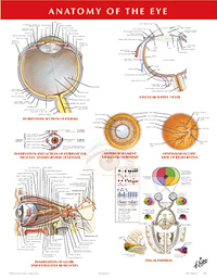
- Page 1.155: Eyeball: Cross Section
- Page 1.156: Anterior and Posterior Chambers of Eye
- Page 1.157: Lens and Supporting Structures: Sectioned in Frontal Pane
- Page 1.158: Lens and Supporting Structures: Horizontal Section
- Page 1.159: Lens and Supporting Structures
- Page 1.16: Intrinsic Arteries and Veins of Eye: Horizontal Section
- Page 1.161: Intrinsic Arteries and Veins of Eye: Ophthalmoscopic Section
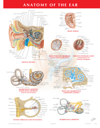
- Page 1.162: Pathway of Sound Reception: Schematic Frontal Section
- Page 1.163: External Ear: Right Auricle
- Page 1.164: External Ear: Right Tympanic Membrane - Viewed Through Speculum
- Page 1.165: Tympanic Cavity: Viewed From External Acoustic Meatus
- Page 1.166: External Ear and Tympanic Cavity: Coronal Oblique Section
- Page 1.167: Tympanic Cavity: Auditory Ossicles
- Page 1.168: Tympanic Cavity: Lateral Wall - Medial View
- Page 1.169: Tympanic Cavity: Medial Wall - Lateral View
- Page 1.17: Right Bony Labyrinth: Anterolateral View - Surrounding Cancellous Bone Removed
- Page 1.171: Right Bony Labyrinth: Dissected - Membranous Labyrinth Remoed
- Page 1.172: Right Membranous Labyrinth with Nerves: Posteromedial View
- Page 1.173: Bony and Membranous Labyrinths: Schema
- Page 1.174: Bony and Membranous Labyrinths: Section Through Turn of Cochlea
- Page 1.175: Orientation of Labyrinth in Skull: Superior Projection
- Page 1.176: Orientation of Labyrinth in Skull: Lateral Projection
- Page 1.177: Pharyngotympanic (Auditory) Tube: Cartilaginous Portion on Skull Base - Inferior View
- Page 1.178: Pharyngotympanic (Auditory) Tube: Section Through Cartilaginous - Tube Closed
- Page 1.179: Pharyngotympanic (Auditory) Tube: Section Through Cartilaginous - Tube Open
- Page 1.18: Meninges and Diploic Veins: Coronal Dissection
- Page 1.181: Diploic and Emissary Veins of Skull
- Page 1.182: Meningeal Arteries
- Page 1.183: Meningeal Arteries
- Page 1.184: Meninges and Superficial Cerebral Veins: Frontal Section
- Page 1.185: Meninges and Superficial Cerebral Veins: Dura Mater Lifted
- Page 1.186: Dural Venous Sinuses: Sagittal Section
- Page 1.187: Dural Venous Sinuses: Cranial Floor - Superior View
- Page 1.188: Dural Venous Sinuses: Coronal Section Through Cavernous Sinus
- Page 1.189: Cerebrum: Lateral View
- Page 1.19: Cerebrum: Lateral View
- Page 1.191: Cerebrum: Insula (Island of Reid) - Lateral View
- Page 1.192: Cerebrum: Brain In Situ - Sagittal Section (Medial View)
- Page 1.193: Cerebrum: Hemisphere with Brainstem Excised - Medial View
- Page 1.194: Cerebrum: Inferior View
- Page 1.195: Ventricles of Brain: Left Lateral Phantom View
- Page 1.196: Ventricles of Brain: Coronal Section - Posterior View
- Page 1.197: Circulation of Cerebrospinal Fluid
- Page 1.198: Basal Nuclei (Ganglia): Horizontal Sections through Cerebrum
- Page 1.199: Basal Nuclei (Ganglia): Schema - Left Lateral View
- Page 1.2: Thalamus
- Page 1.201: Thalamus: Schematic Section
- Page 1.202: Thalamus: Schematic Representation of Thalamus
- Page 1.203: Hippocampus and Fornix: Superior Dissection
- Page 1.204: Hippocampus and Fornix: Schema of Fornix
- Page 1.205: Hippocampus and Fornix: Coronal Section - Posterior View
- Page 1.206: Cerebellum: Superior View
- Page 1.207: Cerebellum: Inferior Surface
- Page 1.208: Cerebellum: Section in Plane of Superior Cerebellar Peduncle
- Page 1.209: Brainstem: Posterolateral View
- Page 1.21: Brainstem: Anterior View
- Page 1.211: Fourth Ventricle and Cerebellum: Posterior View
- Page 1.212: Fourth Ventricle and Cerebellum: Median Sagittal Section
- Page 1.213: Cranial Nerve Nuclei in Brainstem: Schema - Posterior Phantom View
- Page 1.214: Cranial Nerve Nuclei in Brainstem: Schema - Medial Dissection
- Page 1.215: Cranial Nerves (Motor and Sensory Distribution): Schema
- Page 1.216: Olfactory Nerve (I): Schema
- Page 1.217: Optic Nerve (II) (Visual Pathway): Schema
- Page 1.218: Optic Nerve (II): Structure of Retina - Schema
- Page 1.219: Oculomotor, Trochlear, and Abducent Nerves (III, IV, VI): Schema
- Page 1.22: Oculomotor, Trochlear, and Abducent Nerves (III, IV, VI): Schema - Enlargement of Retrobulbar Portions
- Page 1.221: Trigeminal Nerve (V): Schema
- Page 1.222: Trigeminal Nerve (V): Schema - Enlargement of Proximal Portions
- Page 1.223: Facial Nerve (VII): Schema
- Page 1.224: Facial Nerve (VII): Schema - Enlargement of Proximal Portions
- Page 1.225: Vestibulocochlear Nerve (VIII): Schema
- Page 1.226: Glossopharyngeal Nerve (IX): Schema
- Page 1.227: Vagus Nerve (X): Schema
- Page 1.228: Vagus Nerve (X): Schema - Enlargement of Proximal Portions
- Page 1.229: Vagus Nerve (X): Schema - Enlargement of Distal Portions
- Page 1.23: Accessory Nerve (XI): Schema
- Page 1.231: Hypoglossal Nerve (XII): Schema
- Page 1.232: Cervical Plexus: Schema
- Page 1.233: Autonomic Nerves in Neck
- Page 1.234: Autonomic Nerves in Head
- Page 1.235: Ciliary Ganglion: Schema
- Page 1.236: Pterygopalatine and Submandibular Ganglia: Schema
- Page 1.237: Otic Ganglion: Schema
- Page 1.238: Taste Pathways: Schema
- Page 1.239: Arteries to Brain and Meninges
- Page 1.24: Arteries to Brain: Schema
- Page 1.241: Arteries of Brain: Inferior View
- Page 1.242: Arteries of Brain: Inferior View
- Page 1.243: Cerebral Arterial Circle (Willis): Vessels Dissected Out - Inferior View
- Page 1.244: Cerebral Arterial Circle (Willis): Vessels In Situ - Inferior View
- Page 1.245: Arteries of Brain: Frontal View
- Page 1.246: Arteries of Brain: Frontal Section
- Page 1.247: Arteries of Brain: Lateral View
- Page 1.248: Arteries of Brain: Medal View
- Page 1.249: Arteries of Posterior Cranial Fossa
- Page 1.25: Veins of Posterior Cranial Fossa
- Page 1.251: Deep Veins of Brain: Dissection - Superior View
- Page 1.252: Deep Veins of Brain: Dissection - Inferior View
- Page 1.253: Subependymal Veins of Brain
- Page 1.254: Hypothalamus: Principal Nuclei
- Page 1.255: Adenohypophysis and Neurohypophysis
- Page 1.256: Arteries and Veins of Hypothalamus and Hypophysis
- Page 1.257: Head Scans: Sagittal MR Images
- Page 1.258: Head Scans: Axial CT Images
- Page 1.259: Head Scans: Coronal CT Images
- Page 2.001: Back
- Page 2.002: Vertebral Column
- Page 2.003: Thoracic Vertebra (T6): Superior View
- Page 2.004: Thoracic Vertebra (T6): Lateral View
- Page 2.005: Thoracic Vertebra (T12): Lateral View
- Page 2.006: Thoracic Vertebra (T 7-9): Assembled - Posterior View
- Page 2.007: Lumbar Vertebrae (L2): Superior View
- Page 2.008: Lumbar Intervertebral Disc
- Page 2.009: Lumbar Vertebra (L 3-4): Assembled - Posterior View
- Page 2.01: Lumbar Vertebrae (L1-5): Left Lateral View
- Page 2.011: Lumbar Vertebrae: Anteroposterior Radiograph
- Page 2.012: Lumbar Vertebrae: Lateral Radiograph
- Page 2.013: Sacrum and Coccyx: Pelvic Surface
- Page 2.014: Sacrum and Coccyx: Median Sagittal Section
- Page 2.015: Sacrum: Coronal Section through S2 Foramina
- Page 2.016: Sacrum and Coccyx: Dorsal Surface
- Page 2.017: Vertebral Ligaments of Lumbar Region: Left Lateral View - Partially Sectioned in Median Plane
- Page 2.018: Vertebral Ligaments of Lumbar Region: Anterior Vertebral Segments - Posterior View
- Page 2.019: Vertebral Ligaments of Lumbar Region: Posterior Vertebral Segments - Anterior View
- Page 2.02: Vertebral Ligaments of Lumbosacral Region: Left Lateral View
- Page 2.021: Vertebral Ligaments of Lumbosacral Region: Posterior View
- Page 2.022: Spinal Cord and Ventral Rami In Situ
- Page 2.023: Spinal Cord In Situ: Enlargement of Cauda Equina
- Page 2.024: Relation of Spinal Nerve Roots to Vertebrae
- Page 2.025: Relation of Spinal Nerve Roots to Vertebrae: Lateral Protrusion
- Page 2.026: Relation of Spinal Nerve Roots to Vertebrae: Medial Protrusion
- Page 2.027: Motor Impairment Related to Level of Spinal Cord Injury
- Page 2.028: Lumbar Disc Herniation: Clinical Manifestations
- Page 2.029: Lumbar Disc Herniation: Clinical Manifestations
- Page 2.03: Dermatomes
- Page 2.031: Spinal Cord Cross Sections: Sections Through Spinal Cord at Various Levels
- Page 2.032: Spinal Cord Cross Section: Principal Fiber Tracts
- Page 2.033: Autonomic Nervous System: General Topography
- Page 2.034: Autonomic Nervous System: Enlargement of Supradiaphragmatic Portion
- Page 2.035: Autonomic Nervous System: Enlargement of Subdiaphragmatic Portions
- Page 2.036: Autonomic Nervous System: Schema
- Page 2.037: Cholinergic and Adrenergic Synapses: Schema
- Page 2.038: Spinal Membranes and Nerve Roots: Posterior View
- Page 2.039: Spinal Membranes and Nerve Roots: Anterior View
- Page 2.04: Spinal Nerve Origin: Cross Section Through Thoracic Vertebra
- Page 2.041: Spinal Nerve Origin: Section Through Lumbar Vertebra
- Page 2.042: Arteries of Spinal Cord: Schema
- Page 2.043: Arteries of Spinal Cord: Intrinsic Distribution - Thoracic Section
- Page 2.044: Arteries of Spinal Cord: Intrinsic Distribution - Schema
- Page 2.045: Veins of Vertebral Column: Sagittal Section (Without Spinal Cord)
- Page 2.046: Veins of Vertebral Column: Superior View (Without Spinal Cord)
- Page 2.047: Veins of Spinal Cord: Superior View (Magnified With Cord)
- Page 2.048: Muscles of Back: Superficial Layers
- Page 2.049: Muscles of Back: Intermediate Layers
- Page 2.05: Muscles of Back: Deep Layers
- Page 2.051: Nerves of Back
- Page 2.052: Suboccipital Triangle
- Page 2.053: Lumbar Region of Back: Cross Section
- Page 2.054: Typical Thoracic Spinal Nerve
- Page 3.001: Thorax
- Page 3.002: Mammary Gland
- Page 3.003: Mammary Gland: Sagittal Section
- Page 3.004: Arteries of Mammary Gland
- Page 3.005: Lymph Vessels and Nodes of Mammary Gland
- Page 3.006: Bony Framework of Thorax: Anterior View
- Page 3.007: Bony Framework of Thorax: Posterior View
- Page 3.008: Left 1st and 2nd Ribs: Superior View
- Page 3.009: Sternocostal Articulations: Anterior View
- Page 3.01: Middle Rib: Posterior View
- Page 3.011: Costovertebral Joints: Left Lateral View
- Page 3.012: Costovertebral Joints: Transverse Section - Superior View
- Page 3.013: Costovertebral Joints: Right Posterolateral View
- Page 3.014: Cervical Ribs and Related Anomalies 1
- Page 3.015: Cervical Ribs and Related Anomalies 2
- Page 3.016: Cervical Ribs and Related Anomalies 3
- Page 3.017: Anterior Thoracic Wall
- Page 3.018: Anterior Thoracic Wall: Deep Dissection
- Page 3.019: Anterior Thoracic Wall: Internal View
- Page 3.02: Deep Posterior Thoracic Wall
- Page 3.021: Lateral Thoracic Wall
- Page 3.022: Posterior Thoracic Wall
- Page 3.023: Intercostal Nerves and Arteries
- Page 3.024: Diaphragm: Thoracic Surface
- Page 3.025: Diaphragm: Abdominal Surface
- Page 3.026: Phrenic Nerve In Situ
- Page 3.027: Phrenic Nerve: Schema
- Page 3.028: Muscles of Respiration
- Page 3.029: Topography of Lungs: Anterior View
- Page 3.03: Topography of Lungs: Posterior View
- Page 3.031: Lungs In Situ: Anterior View
- Page 3.032: Right Lung: Medial View
- Page 3.033: Left Lung: Medial View
- Page 3.034: Bronchopulmonary Segments: Anterior View
- Page 3.035: Bronchopulmonary Segments: Posterior View
- Page 3.036: Bronchopulmonary Segments: Lateral View
- Page 3.037: Bronchopulmonary Segments: Medial View
- Page 3.038: Trachea and Major Bronchi: Anterior View
- Page 3.039: Trachea: Cross Section
- Page 3.04: Nomenclature of Bronchi: Schema
- Page 3.041: Intrapulmonary Airways: Schema
- Page 3.042: Intrapulmonary Blood Circulation: Schema
- Page 3.043: Great Vessels of Superior Mediastinum
- Page 3.044: Bronchial Arteries
- Page 3.045: Bronchial Arteries: Variations
- Page 3.046: Bronchial Veins
- Page 3.047: Lymph Vessels and Nodes of Lung
- Page 3.048: Autonomic Nerves in Thorax
- Page 3.049: Innervation of Tracheobronchial Tree: Schema
- Page 3.05: Heart In Situ
- Page 3.051: Heart: Anterior Exposure
- Page 3.052: Radiograph of Chest
- Page 3.053: Base of Heart: Posterior View
- Page 3.054: Heart: Diaphragmatic Surface - Posteroinferior View
- Page 3.055: Pericardial Sac: Heart Drawn Out - Left Lateral View
- Page 3.056: Pericardial Sac: Heart Removed - Anterior View
- Page 3.057: Coronary Arteries and Cardiac Veins: Sternocostal Surface
- Page 3.058: Coronary Arteries and Cardiac Veins: Diaphragmatic Surface
- Page 3.059: Coronary Arteries and Cardiac Veins: Variations
- Page 3.06: Coronary Arteries and Cardiac Veins: Variations
- Page 3.061: Right Coronary Artery: Arteriographic View 1
- Page 3.062: Right Coronary Artery: Arteriographic View
- Page 3.063: Left Coronary Artery: Arteriographic View
- Page 3.064: Left Coronary Artery: Arteriographic View
- Page 3.065: Opened Right Atrium: Right Lateral View
- Page 3.066: Opened Right Ventricle: Anterior View
- Page 3.067: Left Ventricle: Flap Opened in Posterolateral Wall
- Page 3.068: Left Atrium and Ventricle: Sectioned With Mitral Valve Cut Away
- Page 3.069: Valves of Heart in Diastole
- Page 3.07: Valves of Heart in Systole
- Page 3.071: Aortic Valve
- Page 3.072: Tricuspid (Right Atrioventricular) Valve
- Page 3.073: Mitral (Left Atrioventricular) Valve
- Page 3.074: Atria, Ventricles and Interventricular Septum
- Page 3.075: Conducting System of Heart: Right Side
- Page 3.076: Conducting System of Heart: Left Side
- Page 3.077: Nerves of Heart
- Page 3.078: Innervation of Heart: Schema
- Page 3.079: Innervation of Blood Vessels: Schema
- Page 3.08: Prenatal Circulation
- Page 3.081: Postnatal Circulation
- Page 3.082: Mediastinum: Right Lateral View
- Page 3.083: Mediastinum: Left Lateral View
- Page 3.084: Esophagus In Situ
- Page 3.085: Topography and Constrictions of Esophagus: Schema - Anterior View
- Page 3.086: Topography and Constrictions of Esophagus: Lateral View
- Page 3.087: Musculature of Esophagus 1
- Page 3.088: Musculature of Esophagus 2
- Page 3.089: Pharyngoesophageal Junction
- Page 3.09: Esophagogastric Junction: Coronal Section
- Page 3.091: Esophagogastric Junction: Anterior View
- Page 3.092: Arteries of Esophagus
- Page 3.093: Arteries of Esophagus: Variations
- Page 3.094: Veins of Esophagus
- Page 3.095: Veins of Esophagus: Submucous Venus Plexus
- Page 3.096: Lymph Vessels and Nodes of Esophagus
- Page 3.097: Nerves of Esophagus: Anterior View
- Page 3.098: Nerves of Esophagus: Posterior View
- Page 3.099: Mediastinum: Cross Section
- Page 3.1: Chest Scans: Axial CT Images - Series of Chest Axial CT Images From Superior to Inferior
- Page 4.001: Abdomen
- Page 4.002: Bony Framework of Abdomen
- Page 4.003: Anterior Abdominal Wall: Superficial Dissection
- Page 4.004: Anterior Abdominal Wall: Intermediate Dissection
- Page 4.005: Anterior Abdominal Wall: Intermediate Dissection - Enlargement of Inguinal Region
- Page 4.006: Anterior Abdominal Wall: Deep Dissection
- Page 4.007: Anterior Abdominal Wall: Deep Dissection - Enlargement of Inguinal Region
- Page 4.008: Rectus Sheath: Cross Sections Above Arcuate Line
- Page 4.009: Rectus Sheath: Cross Sections Below Arcuate Line
- Page 4.01: Anterior Abdominal Wall: Internal View
- Page 4.011: Posterolateral Abdominal Wall
- Page 4.012: Posterolateral Abdominal Wall
- Page 4.013: Arteries of Anterior Abdominal Wall
- Page 4.014: Veins of Anterior Abdominal Wall
- Page 4.015: Nerves of Anterior Abdominal Wall
- Page 4.016: Thoracoabdominal Nerves
- Page 4.017: Inguinal Region: Dissection - Anterior View
- Page 4.018: Inguinal Region: Dissection - Posterior (Internal) View
- Page 4.019: Femoral Sheath and Inguinal Canal
- Page 4.02: Femoral Sheath and Inguinal Canal
- Page 4.021: Inguinal Canal and Spermatic Cord
- Page 4.022: Indirect Inguinal Hernia
- Page 4.023: Posterior Abdominal Wall: Internal View
- Page 4.024: Arteries of Posterior Abdominal Wall
- Page 4.025: Veins of Posterior Abdominal Wall
- Page 4.026: Lymph Vessels and Nodes of Posterior Abdominal Wall
- Page 4.027: Nerves of Posterior Abdominal Wall
- Page 4.028: Quadrants of Abdomen
- Page 4.029: Regions and Planes of Abdomen
- Page 4.03: Greater Omentum and Abdominal Viscera
- Page 4.031: Greater Omentum and Abdominal Viscera: Omentum Raised
- Page 4.032: Mesenteric Relations of Intestines: Transverse Colon Elevated
- Page 4.033: Mesenteric Relations of Intestines: Suspensory Muscle of Duodenum
- Page 4.034: Mesenteric Relations of Intestines: Small Intestine Removed
- Page 4.035: Mesenteric Relations of Intestines: Sigmoid Colon Reflected
- Page 4.036: Omental Bursa: Stomach Reflected
- Page 4.037: Omental Bursa: Cross Section
- Page 4.038: Peritoneum of Posterior Abdominal Wall
- Page 4.039: Stomach In Situ
- Page 4.04: Stomach: Variations in Position and Contour
- Page 4.041: Mucosa of Stomach
- Page 4.042: Mucosa of Stomach: Histology
- Page 4.043: Musculature of Stomach
- Page 4.044: Musculature of Stomach
- Page 4.045: Duodenum In Situ
- Page 4.046: Mucosa of Duodenum
- Page 4.047: Musculature of Duodenum: Layers of Wall
- Page 4.048: Barium Radiograph of Stomach, Duodenum, and Proximal Jejunum
- Page 4.049: Mucosa and Musculature of Jejunum
- Page 4.05: Mucosa and Musculature of Ileum
- Page 4.051: Ileocecal Region
- Page 4.052: Ileocecal Region: Retrocecal Recess Exposed
- Page 4.053: Ileocecal Region: Variations in Posterior Peritoneal Attachment
- Page 4.054: Ileocecal Region: Labial Form of Ileocecal Sphincter
- Page 4.055: Ileocecal Region: Papillary Form of Ileocecal Sphincter
- Page 4.056: Ileocecal Region: Schema of Muscle Fibers at Ileal Orifice
- Page 4.057: (Vermiform) Appendix: Variations in Position
- Page 4.058: (Vermiform) Appendix: Fixed Retrocecal
- Page 4.059: (Vermiform) Appendix: Cross Section
- Page 4.06: Mucosa and Musculature of Large Intestine
- Page 4.061: Sigmoid Colon: Variations in Position
- Page 4.062: Sigmoid Colon: Variations in Position
- Page 4.063: Topography of Liver: Anterior and Posterior Views
- Page 4.064: Topography of Liver: Lateral View
- Page 4.065: Surfaces and Bed of Liver: Anterior View
- Page 4.066: Surfaces and Bed of Liver: Visceral Surface
- Page 4.067: Surfaces and Bed of Liver: Posterior View and Bed of Liver
- Page 4.068: Liver In Situ
- Page 4.069: Variations in Form of Liver
- Page 4.07: Hepatic Portal Segments and Lobules: Parietal Surface
- Page 4.071: Hepatic Portal Segments and Lobules: Visceral Surface
- Page 4.072: Destribution of Vessels and Ducts of Liver
- Page 4.073: Intrahepatic Vascular and Duct Systems: Schema
- Page 4.074: Normal Lobular Pattern of Liver
- Page 4.075: Liver Structure: Schema
- Page 4.076: Intrahepatic Biliary System: Schema
- Page 4.077: Intrahepatic Biliary System: Schema
- Page 4.078: Gallbladder and Extrahepatic Bile Ducts
- Page 4.079: Gallbladder and Extrahepatic Bile Ducts: Sectioned
- Page 4.08: Variations in Cystic Ducts
- Page 4.081: Variations in Hepatic Ducts
- Page 4.082: Junction of Bile Duct and Duodenum
- Page 4.083: Junction of Bile Duct and Duodenum: Dissection
- Page 4.084: Junction of Bile Duct and Duodenum: Reconstruction
- Page 4.085: Variations in Union of Bile and Pancreatic Ducts
- Page 4.086: Pancreas In Situ
- Page 4.087: Pancreas: Histology
- Page 4.088: Pancreas
- Page 4.089: Spleen: Visceral and Diaphragmatic Surfaces
- Page 4.09: Spleen: Cross Section
- Page 4.091: Spleen In Situ
- Page 4.092: Arteries of Stomach, Liver and Spleen
- Page 4.093: Arteries of Stomach, Duodenum, Pancreas, and Spleen
- Page 4.094: Arteries of Liver, Pancreas, Duodenum, and Spleen
- Page 4.095: Celiac Arteriogram
- Page 4.096: Arteries of Duodenum and Head of Pancreas: Reflected to Left
- Page 4.097: Arteries of Duodenum and Head of Pancreas: Posterior View
- Page 4.098: Arteries of Small Intestine
- Page 4.099: Arteries of Large Intestine
- Page 4.1: Collateral Arterial Supply of Liver
- Page 4.101: Variations in Colic Arteries 1
- Page 4.102: Variations in Colic Arteries 2
- Page 4.103: Variations in Colic Arteries 3
- Page 4.104: Variations in Colic Arteries 4
- Page 4.105: Variations in Colic Arteries 5
- Page 4.106: Veins of Stomach, Duodenum, and Pancreas
- Page 4.107: Veins of Stomach, Duodenum, and Pancreas
- Page 4.108: Veins of Small Intestine: Transverse Colon Cut
- Page 4.109: Veins of Small Intestine: Transverse Colon Elevated
- Page 4.11: Veins of Small and Large Intestine
- Page 4.111: Hepatic Portal Vein Tributaries: Portocaval Anastomoses
- Page 4.112: Typical Arrangement of Hepatic Portal Vein
- Page 4.113: Variations of Hepatic Portal Vein
- Page 4.114: Variations of Hepatic Portal Vein
- Page 4.115: Lymph Vessels and Nodes of Stomach
- Page 4.116: Lymph Vessels and Nodes of Stomach: Stomach Reflected
- Page 4.117: Lymph Vessels and Nodes of Small Intestine
- Page 4.118: Lymph Vessels and Nodes of Large Intestine
- Page 4.119: Lymph Vessels and Nodes of Large Intestine
- Page 4.12: Lymph Vessels and Nodes of Pancreas
- Page 4.121: Lymph Vessels and Nodes of Pancreas: Posterior View
- Page 4.122: Autonomic Nerves and Ganglia of Abdomen
- Page 4.123: Nerves of Stomach and Duodenum
- Page 4.124: Nerves of Stomach and Duodenum: View with Stomach Reflected
- Page 4.125: Nerves of Stomach and Duodenum: Hiatal Region
- Page 4.126: Innervation of Stomach and Duodenum: Schema
- Page 4.127: Nerves of Small Intestine
- Page 4.128: Nerves of Large Intestine
- Page 4.129: White Rami Communicantes: Left Side of Schema
- Page 4.13: Innervation of Small and Large Intestines: Right Side of Schema
- Page 4.131: Autonomic Reflex Pathways: Schema
- Page 4.132: Intrinsic Autonomic Plexuses of Intestine: Schema
- Page 4.133: Common Area of Referred Pain in Biliary Disease
- Page 4.134: Innervation of Liver and Biliary Tract: Schema
- Page 4.135: Innervation of Liver: Nerve Fibers Around Fine Branch of Hepatic Artery
- Page 4.136: Common Areas of Pancreatic Pain
- Page 4.137: Innervation of Pancreas: Schema
- Page 4.138: Schema of Intrinsic Nerve Supply of Pancreas
- Page 4.139: Kidneys In Situ: Anterior Views
- Page 4.14: Kidneys In Situ: Anterior Relations of Kidneys
- Page 4.141: Kidneys In Situ: Posterior View
- Page 4.142: Kidneys: Posterior Relations of Kidneys
- Page 4.143: Anterior Surface of Right Kidney
- Page 4.144: Right Kidney Sectioned in Several Planes
- Page 4.145: Suprarenal Gland and Lobulated Kidney of Infant
- Page 4.146: Renal Artery and Vein In Situ
- Page 4.147: Intrarenal Arteries: Frontal Section of Left Kidney - Anterior View
- Page 4.148: Vascular Renal Segments
- Page 4.149: Variations in Renal Artery
- Page 4.15: Variations in Renal Vein
- Page 4.151: Variations in Renal Vein
- Page 4.152: Nephron and Collecting Tubule: Schema
- Page 4.153: Blood Vessels in Parenchyma of Kidney: Schema
- Page 4.154: Ureters in Male: Anterior View
- Page 4.155: Diagonal Course of Ureter through Bladder Wall
- Page 4.156: Ureters in Female: Superior View
- Page 4.157: Arteries of Ureters and Urinary Bladder
- Page 4.158: Lymph Vessels and Nodes of Kidneys and Urinary Bladder
- Page 4.159: Nerves of Kidneys, Ureters and Urinary Bladder
- Page 4.16: Innervation of Kidneys and Upper Ureters: Schema
- Page 4.161: Renal Fascia: Transverse Section through 2nd Lumbar Vertebra
- Page 4.162: Renal Fascia: Sagittal Section Through Right Kidney
- Page 4.163: Arteries and Veins of Right Suprarenal Gland In Situ
- Page 4.164: Arteries and Veins of Left Suprarenal Gland In Situ
- Page 4.165: Cross Section Through Suprarenal Gland
- Page 4.166: Nerves of Suprarenal Glands: Dissection
- Page 4.167: Nerves of Suprarenal Glands: Schema
- Page 4.168: Abdominal Wall and Viscera: Median (Sagittal) Section
- Page 4.169: Abdomen: Omental Bursa - Schematic Transverse Section, Level of T12
- Page 4.17: Abdomen: Abdominal Wall and Intestines
- Page 4.171: Abdominal Scans: Axial CT Images
- Page 5.001: Pelvis and Perineum
- Page 5.002: Bones and Ligaments of Pelvis: Median (Sagittal) Section
- Page 5.003: Bones and Ligaments of Pelvis: Lateral View
- Page 5.004: Bones and Ligaments of Pelvis: Posterior View
- Page 5.005: Bones and Ligaments of Pelvis: Anterior View
- Page 5.006: Female Pelvis: Measurements - Anterior View
- Page 5.007: Male Pelvis: Measurements - Anterior View
- Page 5.008: Female Pelvis: Measurements - Inferior View
- Page 5.009: Female Pelvis: Measurements - Sagittal Section
- Page 5.01: Pelvic Diaphragm of Female: Superior View
- Page 5.011: Pelvic Diaphragm of Female: Medial View
- Page 5.012: Pelvic Diaphragm of Female: Inferior View
- Page 5.013: Pelvic Diaphragm of Female: Lateral View
- Page 5.014: Pelvic Diaphragm: Male
- Page 5.015: Pelvic Diaphragm: Male
- Page 5.016: Pelvic Viscera and Perineum: Female
- Page 5.017: Pelvic Viscera and Perineum: Male
- Page 5.018: Pelvic Contents: Female
- Page 5.019: Pelvic Contents: Male
- Page 5.02: Endopelvic Fascia and Potential Spaces
- Page 5.021: Urinary Bladder: Orientation and Supports
- Page 5.022: Urinary Bladder: Female and Male
- Page 5.023: Sphincters
- Page 5.024: Male and Female Cystourethrograms
- Page 5.025: Pelvic Viscera: Female
- Page 5.026: Uterus, Vagina and Supporting Structures
- Page 5.027: Uterus and Adnexa
- Page 5.028: Pelvic Ligaments
- Page 5.029: Uterus: Age Changes and Muscle Pattern
- Page 5.03: Uterus: Variations in Position
- Page 5.031: Ectopic Pregnancy
- Page 5.032: Female Perineum and External Genitalia (Pudendum or Vulva)
- Page 5.033: Female Perineum (Superficial Dissection)
- Page 5.034: Female Perineum and Deep Perineum
- Page 5.035: Perineum and External Genitalia: Superficial Dissection
- Page 5.036: Perineum and External Genitalia (Deeper Dissection)
- Page 5.037: Penis
- Page 5.038: Perineal Spaces
- Page 5.039: Prostate and Seminal Vesicles
- Page 5.04: Urethra
- Page 5.041: Descent of Testis
- Page 5.042: Scrotum and Contents
- Page 5.043: Homologues of External Genitalia
- Page 5.044: Homologues of Internal Genitalia
- Page 5.045: Testis, Epididymis and Ductus Deferens
- Page 5.046: Rectum In Situ: Female and Male
- Page 5.047: Ischioanal Fossae
- Page 5.048: Rectum and Anal Canal
- Page 5.049: Anorectal Musculature
- Page 5.05: External Anal Sphincter Muscle: Perineal Views
- Page 5.051: Actual and Potential Perineopelvic Spaces
- Page 5.052: Pelvic Scans: Sagittal MR Images
- Page 5.053: Arteries of Rectum and Anal Canal
- Page 5.054: Veins of Rectum and Anal Canal
- Page 5.055: Arteries and Veins of Pelvic Organs: Female
- Page 5.056: Arteries and Veins of Testis
- Page 5.057: Arteries and Veins of Pelvis: Female
- Page 5.058: Arteries and Veins of Pelvis: Male
- Page 5.059: Arteries and Veins of Perineum and Uterus
- Page 5.06: Arteries and Veins of Perineum: Male
- Page 5.061: Lymph Vessels and Nodes of Pelvis and Genitalia: Female
- Page 5.062: Lymph Vessels and Nodes of Perineum: Female
- Page 5.063: Lymph Vessels and Nodes of Pelvis and Genitalia: Male
- Page 5.064: Nerves of External Genitalia: Male
- Page 5.065: Nerves of Pelvic Viscera: Male
- Page 5.066: Nerves of Perineum: Male
- Page 5.067: Nerves of Pelvic Viscera: Female
- Page 5.068: Nerves of Perineum and External Genitalia: Female
- Page 5.069: Neuropathways in Parturition
- Page 5.07: Innervation of Female Reproductive Organs: Schema
- Page 5.071: Innervation of Male Reproductive Organs: Schema
- Page 5.072: Innervation of Urinary Bladder and Lower Ureter: Schema
- Page 6.001: Upper Limb
- Page 6.002: Clavicle and Sternoclavicular Joint
- Page 6.003: Humerus and Scapula: Anterior Views
- Page 6.004: Humerus and Scapula: Posterior Views
- Page 6.005: Shoulder: Anteroposterior Radiograph
- Page 6.006: Shoulder (Glenohumeral) Joint
- Page 6.007: Muscles of Shoulder
- Page 6.008: Muscles of Rotator Cuff
- Page 6.009: Scapulohumeral Dissection
- Page 6.01: Axillary Artery and Anastomoses Around Scapula
- Page 6.011: Pectoral, Clavipectoral and Axillary Fasciae
- Page 6.012: Axilla (Dissection): Anterior View
- Page 6.013: Brachial Plexus: Schema
- Page 6.014: Muscles of Arm: Anterior Views
- Page 6.015: Muscles of Arm: Posterior View
- Page 6.016: Brachial Artery In Situ
- Page 6.017: Brachial Artery and Anastomoses Around Elbow
- Page 6.018: Arm: Serial Cross Sections
- Page 6.019: Bones of Elbow
- Page 6.02: Elbow: Radiographs
- Page 6.021: Ligaments of Elbow
- Page 6.022: Bones of Forearm
- Page 6.023: Individual Muscles of Forearm: Rotators of Radius
- Page 6.024: Individual Muscles of Forearm: Extensors of Wrist and Digits
- Page 6.025: Individual Muscles of Forearm: Flexors of Wrist
- Page 6.026: Individual Muscles of Forearm: Flexors of Digits
- Page 6.027: Muscles of Forearm (Superficial Layer): Posterior View
- Page 6.028: Muscles of Forearm (Deep Layer): Posterior View
- Page 6.029: Muscles of Forearm (Superficial Layer): Anterior View
- Page 6.03: Muscles of Forearm (Intermediate Layer): Anterior View
- Page 6.031: Muscles of Forearm (Deep Layer): Anterior View
- Page 6.032: Forearm: Serial Cross Sections
- Page 6.033: Attachments of Muscles of Forearm: Anterior View
- Page 6.034: Attachments of Muscles of Forearm: Posterior View
- Page 6.035: Carpal Bones
- Page 6.036: Movements of Wrist
- Page 6.037: Ligaments of Wrist
- Page 6.038: Ligaments of Wrist
- Page 6.039: Bones of Wrist and Hand
- Page 6.04: Wrist and Hand: Anteroposterior Radiograph
- Page 6.041: Metacarpophalangeal and Interphalangeal Ligaments
- Page 6.042: Wrist and Hand: Superficial Palmar Dissection
- Page 6.043: Wrist and Hand: Deeper Palmar Dissections
- Page 6.044: Flexor Tendons, Arteries and Nerves at Wrist
- Page 6.045: Bursae, Spaces and Tendon Sheaths of Hand
- Page 6.046: Lumbrical Muscles, Bursae, Spaces and Sheaths: Schema
- Page 6.047: Flexor and Extensor Tendons in Fingers
- Page 6.048: Intrinsic Muscles of Hand
- Page 6.049: Arteries and Nerves of Hand: Palmar Views
- Page 6.05: Wrist and Hand: Superficial Radial Dissection
- Page 6.051: Wrist and Hand: Superficial Dorsal Dissection
- Page 6.052: Wrist and Hand: Deep Dorsal Dissection
- Page 6.053: Extensor Tendons at Wrist
- Page 6.054: Fingers
- Page 6.055: Cutaneous Innervation of Wrist and Hand
- Page 6.056: Arteries and Nerves of Upper Limb
- Page 6.057: Musculocutaneous Nerve
- Page 6.058: Median Nerve
- Page 6.059: Ulnar Nerve
- Page 6.06: Radial Nerve in Arm and Nerves of Posterior Shoulder
- Page 6.061: Radial Nerve in Forearm
- Page 6.062: Cutaneous Nerves and Superficial Veins of Shoulder and Arm
- Page 6.063: Cutaneous Nerves and Superficial Veins of Forearm
- Page 6.064: Cutaneous Innervation of Upper Limb
- Page 6.065: Dermatomes of Upper Limb
- Page 6.066: Lymph Vessels and Nodes of Upper Limb
- Page 6.067: CT Angiograms of Arteries of the Upper limb
- Page 7.001: Lower Limb
- Page 7.002: Hip (Coxal) Bone
- Page 7.003: Hip Joint
- Page 7.004: Hip Joint: Anteroposterior Radiograph
- Page 7.005: Femur
- Page 7.006: Bony Attachments of Muscles of Hip and Thigh: Anterior View
- Page 7.007: Bony Attachments of Muscles of Hip and Thigh: Posterior View
- Page 7.008: Muscles of Thigh: Anterior Views
- Page 7.009: Muscles of Thigh: Anterior Views
- Page 7.01: Muscles of Hip and Thigh: Lateral View
- Page 7.011: Muscles of Hip and Thigh: Posterior Views
- Page 7.012: Psoas and Iliacus Muscles
- Page 7.013: Lumbosacral and Coccygeal Plexuses
- Page 7.014: Lumbar Plexus
- Page 7.015: Sacral and Coccygeal Plexuses
- Page 7.016: Arteries and Nerves of Thigh: Anterior Views
- Page 7.017: Arteries and Nerves of Thigh: Anterior Views
- Page 7.018: Arteries and Nerves of Thigh: Posterior View
- Page 7.019: Nerves of Hip and Buttock
- Page 7.02: Arteries of Femoral Head and Neck
- Page 7.021: Thigh: Serial Cross Sections
- Page 7.022: Knee: Lateral and Medial Views
- Page 7.023: Knee: Anterior Views
- Page 7.024: Knee: Interior
- Page 7.025: Knee: Cruciate and Collateral Ligaments
- Page 7.026: Knee: Anteroposterior Radiograph
- Page 7.027: Right Knee: Posterior and Sagittal Views
- Page 7.028: Arteries of Thigh and Knee: Schema
- Page 7.029: Tibia and Fibula
- Page 7.03: Tibia and Fibula
- Page 7.031: Attachments of Muscles of Leg
- Page 7.032: Muscles of Leg (Superficial Dissection): Posterior View
- Page 7.033: Muscles of Leg (Intermediate Dissection): Posterior View
- Page 7.034: Muscles of Leg (Deep Dissection): Posterior View
- Page 7.035: Muscles of Leg (Superficial Dissection): Anterior View
- Page 7.036: Muscles of Leg (Deep Dissection): Anterior View
- Page 7.037: Muscles of Leg: Lateral View
- Page 7.038: Leg: Cross Sections and Fascial Compartments
- Page 7.039: Bones of Foot
- Page 7.04: Bones of Foot
- Page 7.041: Calcaneus
- Page 7.042: Ankle: Radiographs
- Page 7.043: Ligaments and Tendons of Ankle
- Page 7.044: Ligaments and Tendons of Foot: Plantar View
- Page 7.045: Tendon Sheaths of Ankle
- Page 7.046: Muscles of Dorsum of Foot: Superficial Dissection
- Page 7.047: Dorsum of Foot: Deep Dissection
- Page 7.048: Sole of Foot: Superficial Dissection
- Page 7.049: Muscles of Sole of Foot: First Layer
- Page 7.05: Muscles of Sole of Foot: Second Layer
- Page 7.051: Muscles of Sole of Foot: Third Layer
- Page 7.052: Interosseous Muscles and Deep Arteries of Foot
- Page 7.053: Interosseous Muscles of Foot
- Page 7.054: Femoral Nerve and Lateral Cutaneous Nerve of Thigh
- Page 7.055: Obturator Nerve
- Page 7.056: Sciatic Nerve and Posterior Cutaneous Nerve of Thigh
- Page 7.057: Tibial Nerve
- Page 7.058: Common Fibular (Peroneal) Nerve
- Page 7.059: Dermatomes of Lower Limb
- Page 7.06: Superficial Nerves and Veins of Lower Limb: Anterior View
- Page 7.061: Superficial Nerves and Veins of Lower Limb: Posterior View
- Page 7.062: Lymph Vessels and Nodes of Lower Limb
- Page 7.063: Key Figure for Cross Sections
- Page 8.001: Thorax: Brachiocephalic Vessels
- Page 8.002: Thorax: Aortic and Azygos Arches
- Page 8.003: Thorax: Pulmonary Trunk
- Page 8.004: Thorax: Four Chambers of Heart
- Page 8.005: Abdomen: Esophagogastric Junction
- Page 8.006: Abdomen: Pylorus and Body of Pancreas
- Page 8.007: Abdomen: Origins of Celiac Artery and Portal Vein
- Page 8.008: Abdomen: Renal Hilum and Vessels
- Page 8.009: Abdomen: Ileocecal Junction
- Page 8.01: Abdomen: Sacral Promontory
- Page 8.011: Male Pelvis: Bladder-Prostate Junction
- Page 8.012: Thorax: Heart, Ascending Aorta
- Page 8.013: Thorax: Tracheal Bifurcation, Left Atrium