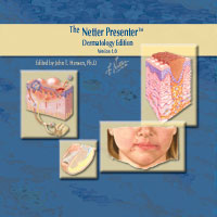
The Netter Presenter Dermatology Edition
This product is no longer available, but individual images or image sets may be purchased
ISBN: 9784M0205REFA
- Page 1.01: Child with Atopic Dermatitis - Dorsal View
- Page 1.02: Child with Atopic Dermatitis - Frontal View
- Page 1.03: Infant with Atopic Dermatitis
- Page 1.04: Infant with Atopic Dermatitis
- Page 1.05: Postauricular Fissures
- Page 1.06: Postauricular Fissures
- Page 1.07: Adolescent with Flexural Lichenification, Trunk Involvement
- Page 1.08: Nipple Eczema
- Page 1.09: Lymphadenopathy in a Child with Atopic Dermatitis
- Page 1.1: Scalp Dermatitis in a Child (Cradle Cap)
- Page 1.11: Facial Xerosis
- Page 1.12: Conjunctivitis Eyelid Inflammation
- Page 1.13: Flexural Lichenification
- Page 1.14: Excoriations of the Foot
- Page 1.15: Hand Dermatitis
- Page 1.16: Popliteal Lesions in an Infant
- Page 1.17: Adult Patient with Atopic Dermatitis of the Hands
- Page 1.18: Atopic Dermatitis Coexisting With Allergic Contact Dermatitis
- Page 1.19: Palmar Hyperlinearity
- Page 1.2: Lichenification and Cheilitis
- Page 1.21: Atopic Dermatitis of the Scalp
- Page 1.22: Child with Scabies - Dorsal View
- Page 1.23: Child with Scabies - Frontal View
- Page 1.24: Allergic Contact Dermatitis
- Page 1.25: Allergic Contact Dermatitis
- Page 2.01: Seborrheic Dermatitis
- Page 2.02: Perioral Dermatitis
- Page 2.03: Contact Dermatitis
- Page 2.04: Vitiligo (Psoralan Burning)
- Page 2.05: Lichen Simplex (Neurodermatitis)
- Page 2.06: Tinea Facia
- Page 2.07: Asteatotic Eczema
- Page 2.08: Pityriasis Alba
- Page 2.09: Psoriasis of the Scalp
- Page 2.1: Lichen Planus
- Page 2.11: Tinea Corporis
- Page 2.12: Lchtyosis Vulgaris
- Page 2.13: Nummular Eczema
- Page 2.14: Lichen Planus
- Page 2.15: Inverse Psoriasis
- Page 2.16: Postauricular Fissures
- Page 2.17: Postauricular Fissures
- Page 2.18: Nipple Eczema
- Page 2.19: Scalp Dermatitis in a Child (Cradle Cap)
- Page 2.2: Facial Xerosis
- Page 2.21: Conjunctivitis Eyelid Inflammation
- Page 2.22: Hand Dermatitis
- Page 2.23: Lichenification and Cheilitis
- Page 2.24: Child with Scabies - Dorsal View
- Page 2.25: Child with Scabies - Frontal View
- Page 2.26: Allergic Contact Dermatitis
- Page 2.27: Allergic Contact Dermatitis
- Page 2.28: Diagnosis of Tinea Pedis
- Page 2.29: Diagnosis of Tinea Pedis
- Page 3.05: External Genital Infections - Herpes Genitalis
- Page 3.06: Balanitis-Herpes Progenitalis
- Page 3.07: Herpes Zoster - Shingles
- Page 3.08: Dermatomes
- Page 4.01: Diabetic Neuropathy
- Page 4.02: Foot Infections
- Page 4.03: Delayed Posttraumatic Osteomyelitis in Diabetic Patient
- Page 4.04: Recurrent Postoperative Osteomyelitis
- Page 4.05: Acute Anterior Compartment Syndrome
- Page 4.06: Deep Infections of Foot
- Page 4.07: Lesions in Diabetic Foot
- Page 4.08: Clinical Evaluation of Patient with Diabetic Foot Lesion
- Page 4.09: Wagner Classification of Diabetic Foot Lesions
- Page 4.11: Complications of Amputation
- Page 4.12: Superficial Veins and Nerves of Lower Limb
- Page 4.13: Superficial Nerves and Veins of Lower Limb
- Page 4.14: Arteries and Nerves of Thigh: Anterior Views - Superficial Dissections
- Page 4.16: Arteries and Nerves of Thigh (Deep Dissection) Anterior View
- Page 4.17: Arteries and Nerves of Thigh (Deep Dissection) posterior view
- Page 4.18: Muscles of Leg (Superficial Dissection) Posterior View
- Page 4.19: Muscles of Leg (Intermediate Dissection) Posterior View
- Page 4.2: Muscles of Leg (Deep Dissection): Posterior View
- Page 4.21: Muscles of Leg (Superficial Dissection): Anterior View
- Page 4.22: Muscles of Leg (Deep Dissection): Anterior View
- Page 4.23: Tendon Sheaths of Ankle - Lateral View
- Page 4.24: Tendon Sheaths of Ankle - Medial View
- Page 4.25: Muscles of Dorsum of Foot (Superficial Dissection)
- Page 4.26: Dorsum of Foot (Deep Dissection)
- Page 4.27: Sole of Foot (Superficial Dissection)
- Page 4.28: Muscles of Sole of Foot: First Layer
- Page 4.29: Muscles of Sole of Foot: Second Layer
- Page 4.3: Muscles of Sole of Foot: Third Layer
- Page 4.31: Interosseous Muscles and Deep Arteries of Foot (Dorsal View)
- Page 4.32: Interosseous Muscles and Deep Arteries of Foot (Plantar View)
- Page 4.33: Anatomy of the Toenail: Sagittal Section
- Page 4.34: Onychomycosis: Classification By Portals of Entry
- Page 4.35: Clinical Examples of Onychomycoses: Distal and Lateral Subungual Onychomycosis
- Page 4.36: Clinical Examples of Onychomycoses: Proximal Subungual Onychomycosis and Proximal White Subungual Onychomycosis
- Page 4.37: Clinical Examples of Onychomycosis
- Page 4.38: Conditions that Mimic Toenail Fungus
- Page 4.39: Prevention and Management of Onychomycosis
- Page 4.4: Consequences of Untreated Onychomycosis in Patients with Diabetes
- Page 4.41: Cutaneous Effects of Diabetes
- Page 4.42: Management of Infected Ulcers: Prevention and Screening
- Page 4.43: Noninfectious Cutaneous Manifestations in the Lower Extremity
- Page 4.44: Noninfectious Cutaneous Manifestations in the Lower Extremity
- Page 4.45: Noninfectious Cutaneous Manifestations in the Lower Extremity
- Page 4.46: Noninfectious Cutaneous Manifestations in the Lower Extremity
- Page 4.47: Diagnosis of Tinea Pedis
- Page 4.48: Diagnosis of Tinea Pedis: Dermatologic Conditions That May Mimic Tinea Pedis
- Page 5.01: Vascular Response to Injury
- Page 5.02: Skin Wound Repair: Inflammatory Phase
- Page 5.03: Skin Wound Repair: Migratory Phase
- Page 5.04: Skin Wound Repair: Proliferative Phase
- Page 5.05: Skin Wound Repair: Maturation Phase
- Page 5.06: Phagocytosis
- Page 5.07: Phagocytosis
- Page 5.08: Epithelial Repair
- Page 5.09: Axon Regeneration
- Page 5.1: Myelin Formation
- Page 5.1: Peripheral Nerver Repair: Late Phase
- Page 5.11: Peripheral Nerver Repair: Early Phase
- Page 5.12: Bone Repair (Early Phase)
- Page 5.13: Bone Repair (Intermediate Phase)
- Page 5.14: Bone Repair (Late Phase)
- Page 5.15: Impairment of Organ Function by Healing Process
- Page 5.16: Collagen Synthesis
- Page 5.17: Wound Closure
- Page 5.17: Wound Closure: A Poor Method
- Page 5.18: Recurrent Postoperative Osteomyelitis
- Page 5.19: Classification of Open Fractures
- Page 5.2: Open Injury With High Risk for Infection
- Page 5.21: Surgical Management of Open Fractures
- Page 5.22: Open Soft Tissue Wounds
- Page 5.22: Pulmonary Embolism
- Page 5.23: Methods of Wound Closure
- Page 5.23: Pressure Ulcer
- Page 5.24: Evolution of a Pressure Ulcer: Early Superficial Ulceration
- Page 5.25: Evolution of a Pressure Ulcer: Late Superficial Ulceration
- Page 5.26: Evolution of a Pressure Ulcer: Early Deep Ulceration
- Page 5.27: Evolution of a Pressure Ulcer: Late Deep Ulceration
- Page 5.28: Preventive Measures - Pressure Relief
- Page 5.29: Surgical Management of Pressure Ulcers
- Page 5.3: Methods of Closure
- Page 5.31: Methods of Closure
- Page 5.32: Methods of Closure
- Page 5.33: Methods of Closure
- Page 5.34: Skin Flaps Based on Direct Cutaneous Arterial Supply
- Page 5.35: Skin Flaps Based on Direct Cutaneous Arterial Supply
- Page 5.36: Skin Flap Design (Transposition Flap)
- Page 5.37: Skin Flap Design (Rotation Flap)
- Page 5.38: Skin Flap Design (Advancement Flap)
- Page 5.39: Excision of Ischial Pressure Ulcer
- Page 5.4: Thigh Flap with Muscle Interposition for Ischial Pressure Ulcer
- Page 5.41: Excision of a Sacral Pressure Ulcer
- Page 5.42: Rotation Flap for a Sacral Pressure Ulcer
- Page 5.43: Gluteus Muscle Interposition for Ischial and Sarcal Pressure Ulcers
- Page 5.44: Excision of Trochanteric Pressure Ulcer
- Page 5.45: Bipedic Flap for Trochanteric Pressure Ulcer
- Page 5.46: Transposition Thigh Flap for Trochanteric Pressure Ulcer