
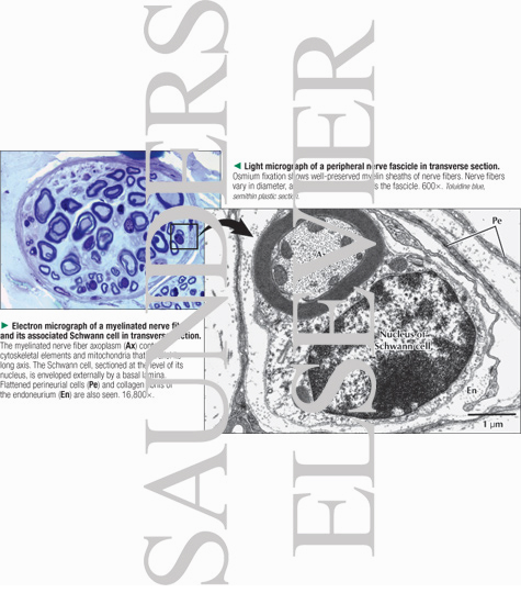
Light Micrograph of a Peripheral Nerve Fascicle In Transverse Section With Electron Micrograph of a Myelinated Nerve Fiber and Its Associated Schwann Cell In Transverse Section
ID: 13456
Title:
Light Micrograph of a Peripheral Nerve Fascicle In Transverse Section With Electron Micrograph of a Myelinated Nerve Fiber and Its Associated Schwann Cell In Transverse Section
Category:
Labeled - Ovalle Histology 1E
Please describe! how you will use this image and then you will be able to add this image to your shopping basket.


 Please Wait...